

Perfusion assessment using transmission laser speckle imaging
a technology of transmission laser and perfusion assessment, applied in the field of perfusion assessment using transmission laser speckle imaging, can solve the problems of pulse oximeters not providing information, pulse oximeters giving erroneous information, or no information at all,
- Summary
- Abstract
- Description
- Claims
- Application Information
AI Technical Summary
Problems solved by technology
Method used
Image

Examples
example 1
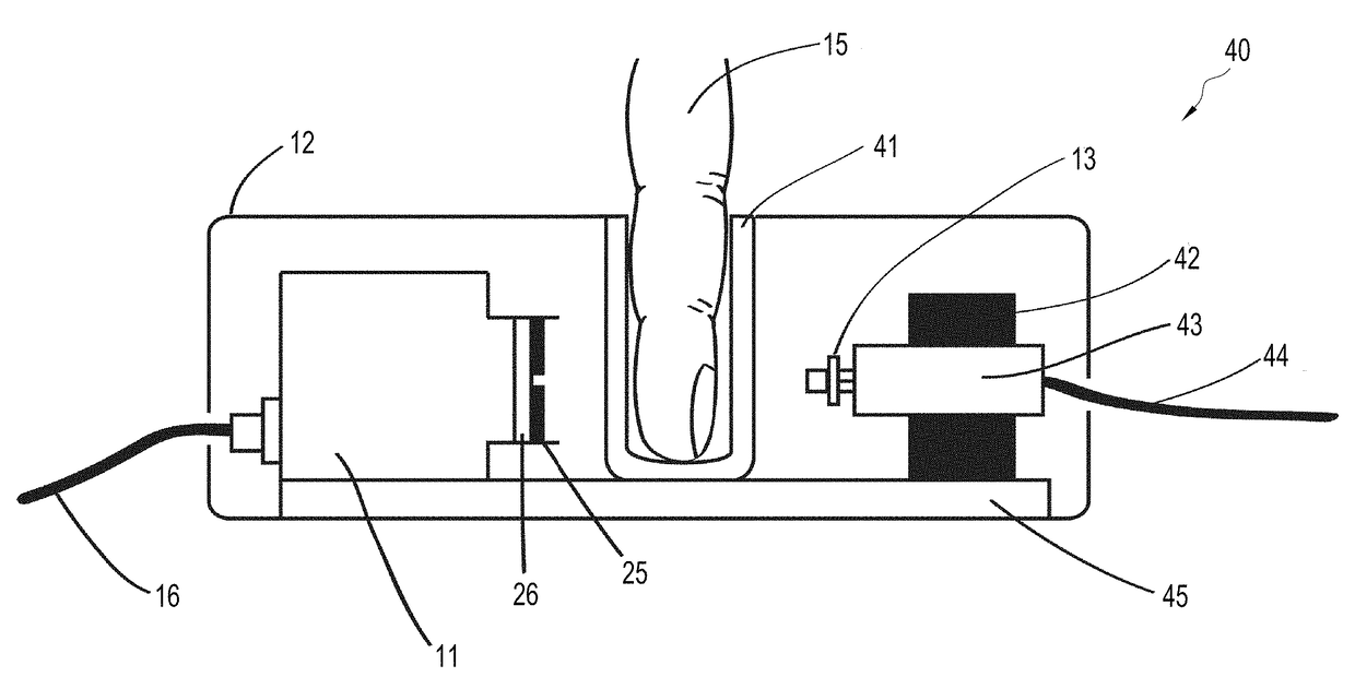
[0079]This example investigated whether the embodiment shown in FIGS. 4-ab (“the FIG. 4 embodiment”) was capable of detecting perfusion changes in the ring finger 15 of a human subject during an arm cuff occlusion. For this experiment, the tip of the patient's ring finger 15 was illuminated using a single mode laser diode 13 (power of 90 mW, wavelength of 789 nm, Roithner LaserTechnik ADL-78901TL, Vienna, Austria). A laser diode driver (Thorlabs Inc. LDC200C, Newton, N.J.) was used to adjust the power output of the laser diode 13. A polydimethylsiloxane (“PDMS”) cylinder 41 had been previously molded around a separate patient's finger. The thickness of the PDMS surrounding the patient's finger was approximately 3 mm. The distance between the finger 15 and laser 13 was 5 mm. The distance between the finger 15 and the detector 11 was 10 mm.
[0080]Light transmitted through the patient's ring finger 15 was then incident on an opaque sheet of aluminum cut into a circle 25 with a diameter ...
example 2
[0092]In this experiment, the FIG. 4 embodiment was used to measure perfusion from a phantom finger in place of a human finger. Blood vessels within the finger-simulating phantom were modeled via the inclusion of Tygon tubing (Norton Performance Plastics 720997, Akron, Ohio). The tubing was wound in a coil ca. 6 mm in diameter up the length of the whole phantom. The tubing has 0.050 inch inner diameter and a 0.090 inch outer diameter. The coiled tubing was covered in a layer PDMS that had been mixed with titanium oxide (Sigma Aldrich T8141-100G, St. Louis, Mo.) at a concentration of 0.74 mg / mL. This provided a PDMS solution with a reduced scattering coefficient of ca. 1 mm−1 at a wavelength of 789 nm. This reduced scattering coefficient was used because it is similar to that found in bulk human tissue.
[0093]To simulate blood flow in the phantom finger, defibrinated sheep blood (Quad Five 610, Ryegate, Mont.) was pumped through the coiled Tygon tubing. Blood was pumped using a perist...
example 3
[0097]The purpose of this experiment was to compare perfusion measurements made using some embodiments of the invention and a laser Doppler flowmetry system (Perimed PeriFlux System 5000, Ardmore, Pa.). Laser Doppler flowmetry was chosen for this comparison because it is very commonly used in the clinic to assess perfusion and is considered a gold standard for doing so. Such assessment is thought to be prognostic of wound healing, flap monitoring, ulcer health, and others.
[0098]In this experiment, the phantom finger described in EXAMPLE 2 was used in place of a patient's finger. The apparatus used to measure perfusion was the same embodiment illustrated in FIGS. 4a-b, except the PDMS finger cavity 41 was removed (“Example 3 embodiment”). A pulsatile pump (LMI Unidose U12-281, FL) was used to pump a 1% solution of Intralipid (Baxter Healthcare Corporation NDC 0338-0519-03, Deerfield, Ill.) diluted with water through the length of tubing. This concentration of Intralipid was used beca...
PUM
 Abstract
Description
Claims
Application Information