Methods and compositions for treating genetically linked diseases of the eye

a technology of genetic linkage and composition, applied in the field of gene therapy, can solve the problems of untreated area being susceptible to retinal detachment, affecting the effect of treatment effect, and deteriorating condition with age, and achieve the effect of minimizing or avoiding unwanted inflammatory responses or other unwanted side effects

Active Publication Date: 2018-01-23
UNITED STATES OF AMERICA
View PDF4 Cites 4 Cited by
  • Summary
  • Abstract
  • Description
  • Claims
  • Application Information

AI Technical Summary

Benefits of technology

[0011]We have surprisingly found that the inventive compositions and methods of administration are capable of inducing the production of proteins in tissues of the eye while minimizing or avoiding unwanted inflammatory responses or other unwanted side effects. Thus, the invention provides expression vectors and therapeutic methods of using such vectors in the treatment of diseases of the eye, particularly disorders of the eye resulting from failure to produce a specific protein in the eye, or the production of a non-functional protein in the eye.

Problems solved by technology

This is evident on clinical electroretinogram (ERG) testing as a characteristic reduction of the b-wave response (from second-order retinal bipolar cells) relative to the photoreceptor a-wave, which frequently gives rise to an ‘electronegative ERG waveform.’ The fragile XLRS retina is more prone to disease related complications, such as vitreous hemorrhage and retinal detachment, and the condition worsens with age.
While the LCA trials used subretinal injection to deliver the vector, this delivery strategy may be problematic for an XLRS trial, as subretinal injection gives geographically localized delivery.
Although this amount of transduction is sufficient to cover the vicinity of the macula, much of the retina would probably not be transduced, and the untreated area would remain susceptible to retinal detachment and vitreous hemorrhage, which are the major causes of vision loss with this disease.
Subretinal injection of retinas with schisis pathology may be challenging and pose a significant risk to the visual function of the subject.
Adhesion of the vitreous to the retina may cause further laminar splitting of the fragile XLRS retina when the surgeon attempts to separate the vitreous from the retina.
In addition, the injection itself may also be difficult.
If the tip of the injection needle is not positioned deep enough, vector solution may be inadvertently routed into the schisis cavities and exacerbate the intraretinal splitting.
However, administration of AAV2 vector leads to a therapy-limiting immune response in the eye, since humans have a high preexisting immunity to AAV2.

Method used


Image

  • Methods and compositions for treating genetically linked diseases of the eye
  • Methods and compositions for treating genetically linked diseases of the eye
  • Methods and compositions for treating genetically linked diseases of the eye

Examples


example 1

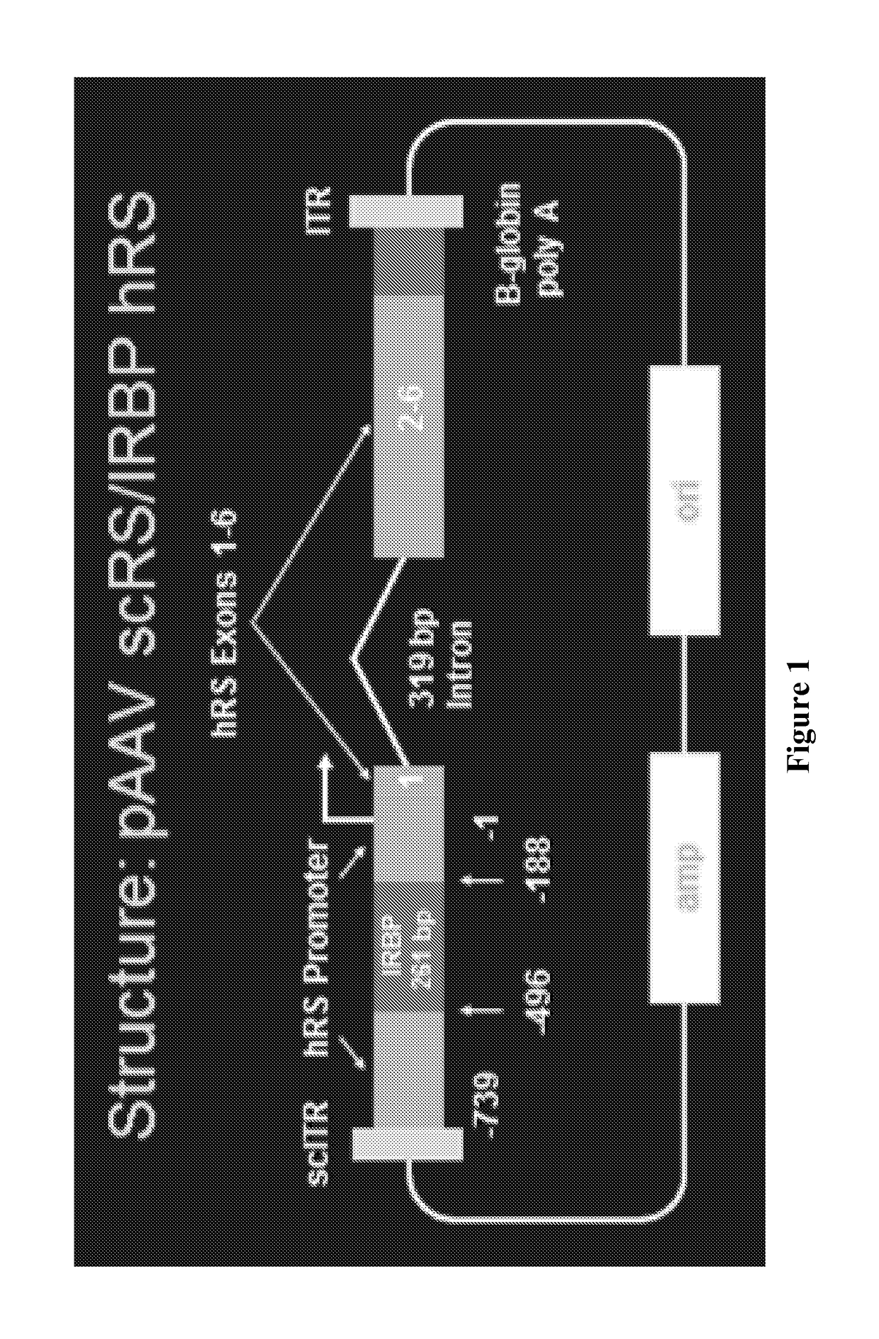
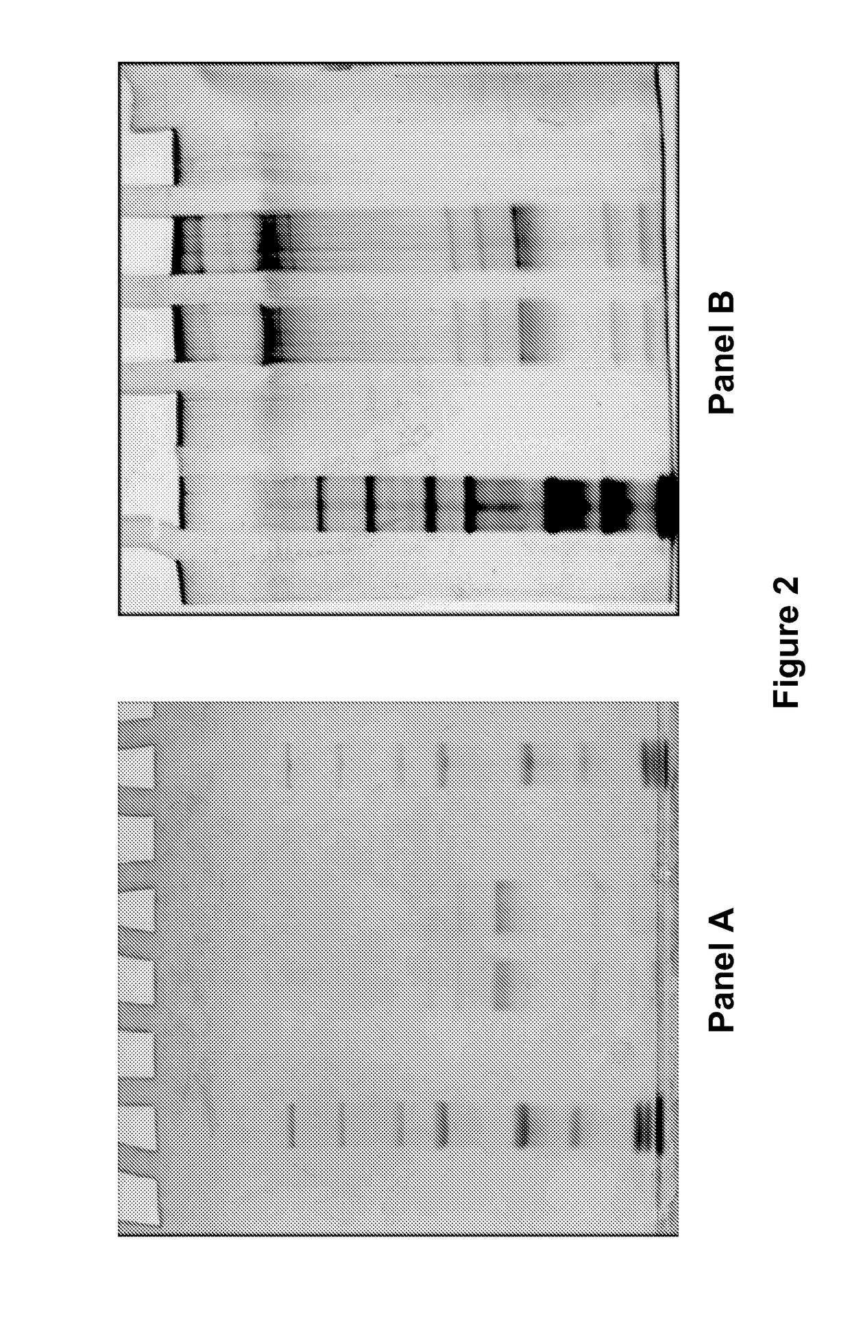
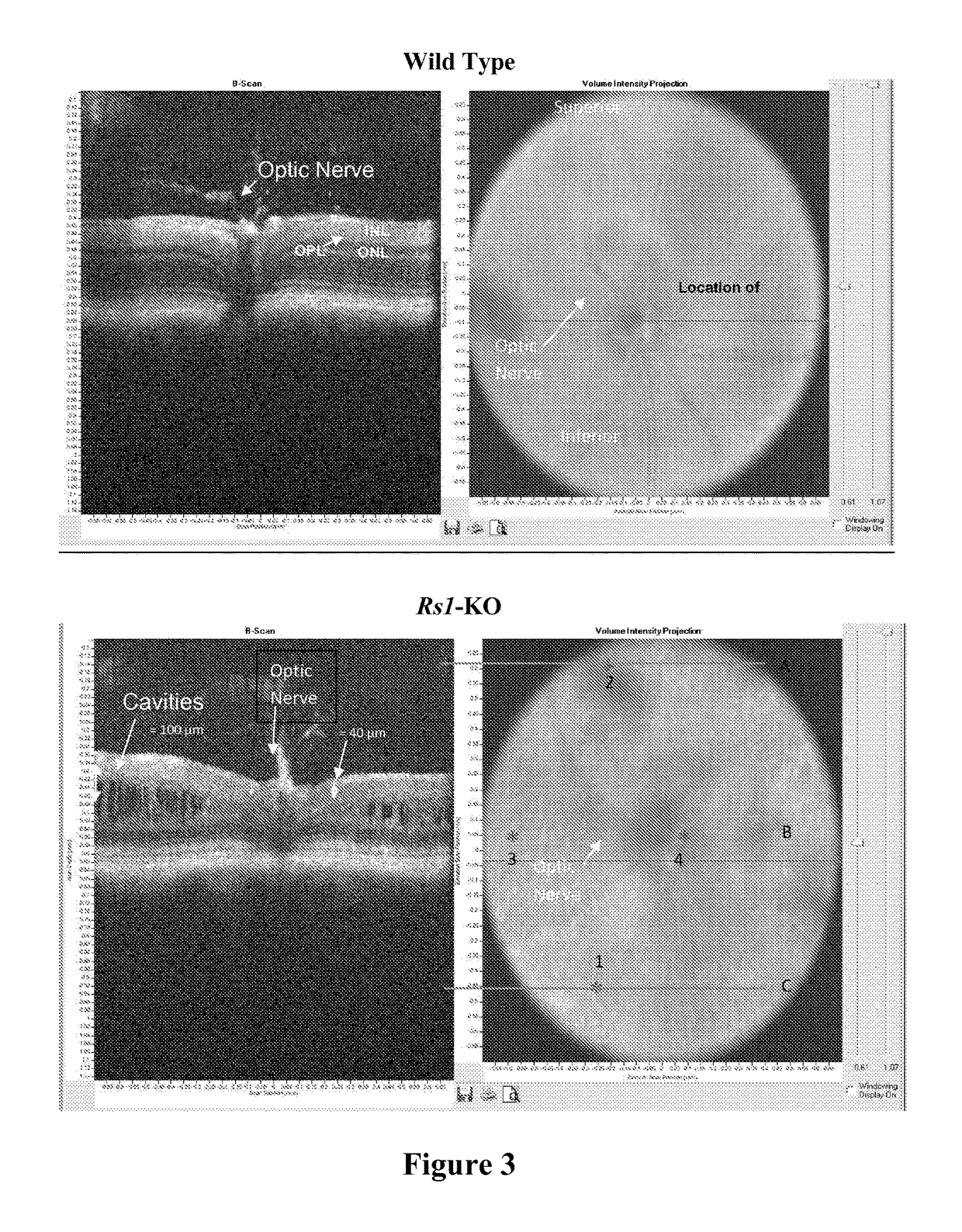
[0100]A study was conducted to evaluate the ability of the proposed clinical adeno-associated virus (AAV) retinoschisin vector, AAV8 scRS / IRBP hRS, to preserve retinal function and structure, and to mediate retinoschisin protein expression when administered intravitreally to the retinoschisin deficient Rs1-KO mouse. AAV8 scRS / IRBP hRS vector at doses of 1.0e6, 1.0e7, 5.0e7, 1.0e8, 5.0e8 and 2.5e9 vg / eye, or vehicle, were administered by intravitreal injection to 18-34 day old Rs1-KO mice. The contralateral eye was not injected. Corneal electroretinogram (ERG) a-wave and b-wave amplitudes were measured at 11 to 15 weeks and 6 to 9 months post injection (PI), retinal cavity formation was measured by optical coherence tomography (OCT) at 12 to 16 weeks PI, and retinoschisin protein expression was measured by immunohistochemistry at 12 to 18 weeks and 6 to 9 months PI. ERG and OCT are used clinically as indicators of retinal function and structure, respectively. At 11-15 weeks PI, eyes ...

example 2

[0162]A study was conducted to assess the tolerability of expression vectors of the invention in rabbit eyes compared with control injections of vehicle alone. Briefly, thirty-nine New Zealand White rabbits (age 6-7 months at injection; weight 2.4-3.8 kg) were used in the present study. All in life procedures were conducted in compliance with the ARVO Statement for the Use of Animals in Ophthalmic and Vision Research and were approved by the Animal Care and use Committee of the National Eye Institute.

[0163]Vectors or vehicle were administered in the right eye by intravitreal injection using ½ cc Insulin Syringes with permanently attached 28 gauge needle (Ultrafine U-100 syringe—BD Biosciences, San Jose, Calif.) in an injection volume of 50 ul. Syringes were loaded under sterile conditions in a laminar flow hood on the day of injection. Rabbits were anesthetized with IM ketamine, 40 mg / kg, and xylazine, 3 mg / kg. Sterile surgical instruments were used and the animals were prepared ase...


PUM

PropertyMeasurementUnit
diameteraaaaaaaaaa
pHaaaaaaaaaa
pHaaaaaaaaaa

Abstract

Expression vectors and therapeutic methods of using such vectors in the treatment of diseases of the eye resulting from failure to produce a specific protein in the eye, or the production of a non-functional protein in the eye.

Description

CROSS REFERENCE TO RELATED APPLICATION[0001]This application is a national stage application under 35 U.S.C. 371 and claims the benefit of International Application No. PCT / US2014 / 016389 having an international filing date of Feb. 14, 2014, which designated the United States, which PCT application claimed the benefit of U.S. Provisional Patent Application Ser. No. 61 / 765,654 filed Feb. 15, 2013 and U.S. Provisional Patent Application Ser. No. 61 / 815,636 filed Apr. 24, 2013, the disclosures of each of which are incorporated herein by reference.TECHNICAL FIELD[0002]The invention relates to gene therapy and specifically, expression vectors and therapeutic methods of using such vectors in the treatment of diseases of the eye resulting from failure to produce a specific protein in the eye, or the production of a non-functional protein in the eye.BACKGROUND OF INVENTION[0003]Several diseases of the eye result from an underlying genetic cause. For example, in some diseases, a mutation in a...

Claims


Application Information

Patent Timeline
Patent Type & Authority Patents(United States)
IPC IPC(8): C12N15/86A61K9/00C07K14/47C12N7/00A61K38/17A61K48/00
CPCC12N15/86A61K9/0046A61K38/1709A61K48/00C07K14/47C12N7/00A61K48/0058C12N2830/42C12N2750/14143C12N2830/008A61P27/02
Inventor SIEVING, PAUL ALBERTBUSH, RONALD AVERYCOLOSI, PETER C.ZENG, YONG
Owner UNITED STATES OF AMERICA
Why Eureka
Try Eureka