Fluorescent molecule fault imaging device

A fluorescent molecular tomography and imaging device technology, which is applied in fluorescence/phosphorescence, diagnosis, material excitation analysis, etc., can solve the problems of poor data set validity, few light source-detector data pairs, and small effective data set, etc., to improve imaging Effects of speed, shorter exposure time, and shorter imaging time

Inactive Publication Date: 2010-04-07
TSINGHUA UNIV
View PDF4 Cites 1 Cited by
  • Summary
  • Abstract
  • Description
  • Claims
  • Application Information

AI Technical Summary

Problems solved by technology

Although this approach yields larger datasets, up to 10 source-detector pairs 6 , but due to factors such as the field of view of the camera, depth of field, exposure time, and the scattering characteristics of biological tissues, only a small area of ​​the fluorescence signal image on the boundary of the small animal can be obtained each time, and the obtained fluorescence signal image can be used There are few light source-detector data pairs, the validity of the data set is poor, and the effective data set is small
In order to obtain an effective large data set of fluorescent signals on the boundary of the experimental small animal sample body, to improve the quality of the reconstructed image, it is necessary to perform exposure imaging at small intervals (imaging once every N degrees), and this results in imaging times M (M=360 / N), it takes a long time to acquire a series of fluorescence images
[0004] In the existing non-contact fluorescent molecular tomography imaging device, the CCD camera directly shoots the experimental small animals, and the available excitation light source-detector data pairs in the obtained fluorescent signal images are small, the effective data set is small, and the system imaging time is relatively small. Long, not only affects the survival of experimental small animal samples, is not conducive to the construction of small animal models in the body for a long time, and the experimental imaging efficiency is low, it is difficult to achieve high-throughput experiments, which limits the application of fluorescence molecular tomography
In conclusion, the above-mentioned fluorescence molecular tomography devices all suffer from some defects.

Method used


Image

  • Fluorescent molecule fault imaging device
  • Fluorescent molecule fault imaging device
  • Fluorescent molecule fault imaging device

Examples


Embodiment Construction

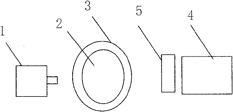
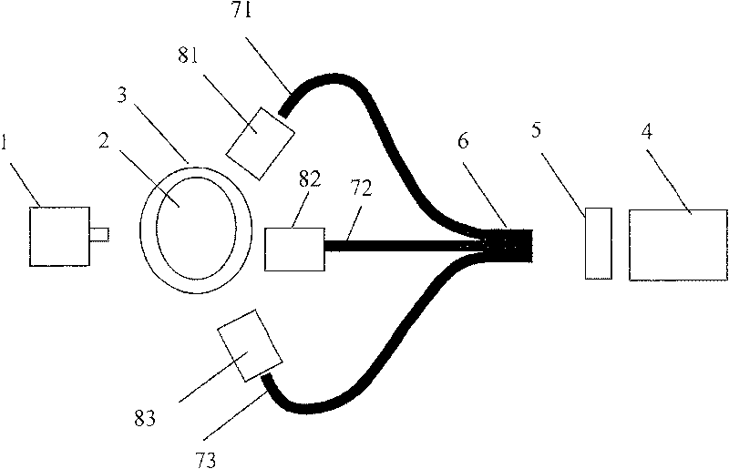
[0020] The present invention is characterized in that it contains: an excitation light source, a filter, a rotary table, an image transmission fiber, an optical lens and a detector, wherein;

[0021] A rotating table, used to drive the experimental small animal sample to rotate;

[0022] An excitation light source, located on one side of the rotary table, is used to emit excitation light to the experimental small animal sample;

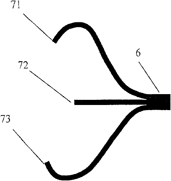
[0023] Image transmission optical fiber, one end is a plurality of forked ends, and the other end is a composite end that merges the plurality of forked ends;

[0024] The number of optical lenses is equal to the number of forked ends of the image-transmitting optical fiber. Each optical lens images the experimental small animal sample on each forked end face of the imaging optical fiber, and the optical lenses are located in a distributed manner. the other side of the experimental small animal sample;

[0025] The detector is a camera, which is aim...


PUM


Abstract

The invention discloses a fluorescence molecule paraclase imaging device, belongs to the technical field of fluorescence molecule paraclase imaging, and is characterized in that: the device comprisesarotating platform that is used for bringing an experimental small animal sample into autorotation, an exciting light source which is arranged at one side of the small animal sample, a plurality of op tical lenses that are arranged at the other side oriented to the experimental small animal sample, an image transmitting optical fiber with each branch end aiming at each optical lens in a non-contactway, a camera with a lens aiming at a composite end of an imaging optical fiber and receiving fluorescence and realizing imaging in a non-contact way, and optical filtering sheets which are respectiv ely arranged in front of each optical lens, used for filtering exciting lights and allowing only fluorescence to enter each optical lens, the optical filtering sheet can be a single sheet arranged infront of the camera. The optical lenses can be arranged in intervals with equal angle Theta and oriented to the experimental small animal sample. The fluorescence molecule paraclase imaging device has the advantages of more exciting light source-detector data pairs, short exposure time, quick imaging speed and the like.

Description

technical field [0001] The invention belongs to the comprehensive technical field of application of near-infrared laser, computer, machinery, electronics and image processing in biomedicine. Background technique [0002] Fluorescence molecular tomography (Fluorescence molecular tomography, also known as fluorescence molecular tomography) uses specific fluorescent molecular probes that have been widely used in biological and medical research to mark specific molecules or cells, and to analyze molecular levels in vivo. In vivo observation of the change of the target, and information such as the distribution of the target is provided through image reconstruction, which overcomes the limitations of planar imaging and obtains more information about biological and medical behaviors. It has the advantages of high sensitivity, quick and easy, low cost, and relatively high throughput. It supports in vivo research on related molecular events and meets the requirements of systematic ob...

Claims


Application Information

Patent Timeline
Patent Type & Authority Patents(China)
IPC IPC(8): A61B5/00G01N21/64
Inventor 白净陈延平汪待发
Owner TSINGHUA UNIV
Why Eureka
Try Eureka