Superconducting probe with bracket

A technology of superconducting and probe brackets, which is applied in catheters, surgery, etc., can solve the problems of unclean abortion, inconvenient clinical application of ultrasonic probes, and large space occupation, and achieve the effect of reducing space occupation

Inactive Publication Date: 2012-05-02
YANGZHOU KELAN ELECTRONICS
View PDF0 Cites 0 Cited by
  • Summary
  • Abstract
  • Description
  • Claims
  • Application Information

AI Technical Summary

Problems solved by technology

[0002] With the continuous improvement and development of ultrasound technology and the application of digital technology in the field of ultrasound, in the current gynecological surgery, ultrasound image monitoring has begun to be used for surgery, but the existing ultrasound probes have many inconveniences and limitations in clinical application sex
For example, a gynecological operation probe combined with a vaginal speculum must be inserted into the vagina after the ultrasonic probe is combined with the outside of the vaginal speculum. As a result, the probe occupies the external space of the speculum in clinical use. During the operation, the expansion of the speculum itself has brought pain to the patient. If the probe is added to the outside of the speculum, the occupied space will be reduced. Larger; for multiparous women, this method can be barely applied, but for caesarean section women or nulliparous women, the use of this technology will bring greater pain to the patient, and the probe and speculum in this method It is relatively fixed, and the detection depth cannot be adjusted back and forth according to the physiological structure of the patient's vagina
Another example is another gynecological operation probe that is used in combination with cervical forceps. In use, the ultrasonic probe and cervical forceps can be combined and then inserted into the vagina through a speculum. Because the probe is combined with cervical forceps, it can be used in clinical operations. During the process, the cervical forceps must be used frequently, which results in sometimes no images, which is not practical and inconvenient in clinical use, and may result in other sequelae such as miscarriage. Due to the large number of clamping parts connected to the cervical forceps, the volume is also large after combination, which will block the normal vision of the surgeon in specific clinical use, bring a lot of inconvenience to the operation process, and fail to achieve the proper monitoring of the entire operation role and effect

Method used


Image

  • Superconducting probe with bracket
  • Superconducting probe with bracket
  • Superconducting probe with bracket

Examples


Embodiment 1

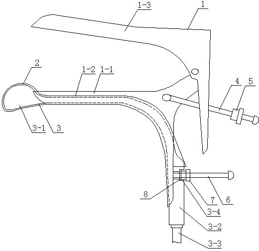
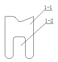
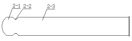
[0018] like figure 1 , 2 , 3, the present invention includes a probe holder 1 and a probe 3 inserted into a patient's vagina in an obstetrics and gynecology operation. The probe holder 1 and the superconducting probe 3 shells are made of metal or plastic, and the probe holder 1 includes an upper wing 1 -3 and the lower wing 1-1, the upper wing 1-3 is hingedly connected with the lower wing 1-1 and positioned by the positioning bolt 4 and the positioning nut 5. The lower wing 1-1 is provided with a notch 1-2, the lower wing 1-1 is fixed with a connecting bolt 6, the bolt 6 is threaded to connect the stepped nut 7, the bolt 6 is set with a T-shaped hollow washer 8, and a T-shaped hollow washer 8 Set between the lower wing 1 - 1 and the stepped nut 7 , the diameter of the end of the stepped nut 7 near the T-shaped hollow washer 8 is smaller than the diameter of the end of the T-shaped hollow washer 8 near the stepped nut 7 . The probe 3 includes a sound head 3-1, a probe handle ...

Embodiment 2

[0021] like Figure 5 As shown, one end of the T-shaped hollow washer 8 close to the stepped nut 7 is arranged outside the stepped groove 3-4. All the other structures are with embodiment 1.

[0022] In clinical use, put the disposable isolation sleeve 2 on the probe 3, so that the front end of the disposable isolation sleeve 2 tightly wraps the acoustic head part 3-1 of the probe 3, and put the probe 3 covered with the disposable isolation sleeve 2 Put it into the inner side of the probe holder 1, and insert the bolt 6 of the handle of the probe holder into the stepped groove 3-4 of the handle of the probe. According to the physiological structure of the patient's vagina, it can be gently pushed forward or pulled backward by hand during specific use. Move the probe 3 to obtain a clear and effective ultrasound image, and then rotate the stepped nut 7 on the bolt 6 at the probe handle 3-2 and insert it into the stepped groove 3-4 at the probe handle 3-2, so that the probe 3 ca...


PUM


Abstract

The invention discloses a superconducting probe with a bracket, relating to a technology of medical instruments for gynecological and obstetrical operations. The superconducting probe with the bracket comprises a probe bracket and a probe. The probe bracket comprises a lower wing; the probe includes a sound head and a probe handle; the back end of the sound head is connected with the front end of the probe handle; a disposable isolating sleeve is sleeved outside the probe; the lower ring is fixedly connected with a bolt, a screw thread on which is connected with a nut; and a T-shaped hollow gasket which is sleeved on the bolt is arranged between the lower wing and the nut. The lower wing is provided with a recess where the probe sleeved with the disposable isolating sleeve is permeated and arranged; a step-shaped groove is arranged at the lower part of the probe handle; and the T-shaped hollow gasket is matched and arranged in the step-shaped groove. By integrally combining the disposable isolating sleeve with the internal side of the probe bracket, the probe disclosed by the invention not only reduces precious space occupation of a speculum, but also can provide a guarantee for accurate positioning of artificial abortion operations.

Description

technical field [0001] The invention relates to a technology of medical equipment for obstetrics and gynecology surgery. Background technique [0002] With the continuous improvement and development of ultrasound technology and the application of digital technology in the field of ultrasound, in the current gynecological surgery, ultrasound image monitoring has begun to be used for surgery, but the existing ultrasound probes have many inconveniences and limitations in clinical application sex. For example, a gynecological operation probe combined with a vaginal speculum must be inserted into the vagina after the ultrasonic probe is combined with the outside of the vaginal speculum. As a result, the probe occupies the external space of the speculum in clinical use. During the operation, the expansion of the speculum itself has brought pain to the patient. If the probe is added to the outside of the speculum, the occupied space will be reduced. Larger; for multiparous women,...

Claims


Application Information

Patent Timeline
Patent Type & Authority Applications(China)
IPC IPC(8): A61B8/12
Inventor 不公告发明人
Owner YANGZHOU KELAN ELECTRONICS
Why Eureka
Try Eureka