
Automatically initialized local active contour model heart and cerebral vessel segmentation method

An active contour model, cardiovascular and cerebrovascular technology, applied in image analysis, image data processing, instruments, etc., can solve problems such as large computational load, uneven gray distribution, and strict requirements for initial contour settings.

Active Publication Date: 2013-03-20
BEIJING NORMAL UNIVERSITY
View PDF2 Cites 19 Cited by
  • Summary
  • Abstract
  • Description
  • Claims
  • Application Information

AI Technical Summary

Problems solved by technology

For the problem of uneven gray distribution of blood vessels, especially small blood vessels, it is difficult to accurately segment
In the process of contour line evolution in the current method, usually all pixels in the image domain participate in the calculation, and the calculation load is large
In addition, the traditional blood vessel segmentation method based on the geometric active contour model is easy to fall into the local minimum, and the setting requirements for the initial contour line are relatively strict.

Method used


Image

  • Automatically initialized local active contour model heart and cerebral vessel segmentation method
  • Automatically initialized local active contour model heart and cerebral vessel segmentation method
  • Automatically initialized local active contour model heart and cerebral vessel segmentation method

Examples


Embodiment Construction

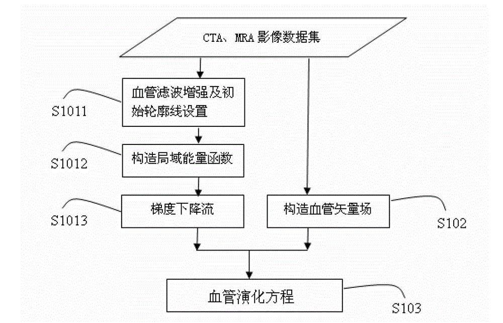
[0044] The present invention will be described in further detail below in conjunction with the accompanying drawings, and the method for cardiovascular and cerebrovascular segmentation based on the local active contour model of the blood vessel shape of the present invention will be described in detail below through the accompanying drawings.

[0045] Due to the small grayscale contrast between blood vessels and surrounding tissues in medical image data, it is difficult to effectively enhance blood vessels by traditional methods such as Gaussian filtering. The point realization of the contour line and the surrounding small neighborhood improves the evolution efficiency; the blood vessel vector field is defined according to the position of the center line of the blood vessel and the direction of the active contour line, and assists the evolution of the active contour line along the direction of the blood vessel. The overall flow of the cardiovascular and cerebrovascular segmenta...


PUM


Abstract

The invention provides an automatically initialized local active contour model heart and cerebral vessel segmentation method. The method comprises the following steps: performing anisotropic filtering on a three-dimensional vessel according to the tubular feature of the vessel in medical image data to acquire an initial contour line; constructing a local energy function; minimizing the local energy function by a gradient descent method to acquire the corresponding gradient descent flow; constructing a vessel vector field according to the vessel shape measurement of the medical image data; and finally combining the gradient descent flow and the vessel vector field to acquire a vessel evolution equation. The method can automatically set the initial contour line according to the vessel shape and is precise in segmentation, high in efficiency and high in robustness.

Description

technical field [0001] The invention relates to the field of computer application technology, in particular to a method for segmenting blood vessels by using a local active contour model and a blood vessel vector field, which is mainly used in the fields of medical teaching and calculation-aided diagnosis and treatment. Background technique [0002] At present, cardiovascular and cerebrovascular diseases have become the "first killer" of human health. The number of people who die from cardiovascular and cerebrovascular diseases in the world is as high as 15 million every year. In China, there are more than 270 million patients with cardiovascular and cerebrovascular diseases, and nearly 3 million people die from cardiovascular and cerebrovascular diseases every year, accounting for 51% of the total number of deaths. Among the surviving patients, 75% lost their ability to work to varying degrees, and 40% were seriously disabled. [0003] Cardiovascular and cerebrovascular di...

Claims


Application Information

Patent Timeline
Patent Type & Authority Applications(China)
IPC IPC(8): G06T7/00
Inventor 田沄周明全武仲科赵世凤王醒策解立志
Owner BEIJING NORMAL UNIVERSITY
Learn More