Medical imaging data streaming

a medical imaging and data technology, applied in the field of medical imaging data streaming, can solve the problems of not being able to find ultrasound imaging specialists in remote or rural areas, not all ultrasound examinations may actually be performed with a doctor in the examination room, and not being able to provide expert doctors or specialists with specific skills in ultrasound examination and diagnosis, etc., to achieve control over the pre-processing

Active Publication Date: 2008-08-26
GE MEDICAL SYST GLOBAL TECH CO LLC
View PDF33 Cites 27 Cited by
  • Summary
  • Abstract
  • Description
  • Claims
  • Application Information

AI Technical Summary

Benefits of technology

[0014]Additionally, the remote specialist may use the remote console controls to transmit commands back to the ultrasound imaging system. The commands may be used to manipulate the pre-processing or post-processing functions of the ultrasound data processor. Thus, the remotely located doctor may exert considerable control over the pre-processing and post-processing functions of the ultrasound data processor.

Problems solved by technology

However, not all ultrasound examinations may actually be performed with a doctor in the examination room.
In today's highly specialized medical society, expert doctors or specialists with skills in specific fields such as ultrasound examination and diagnosis for example, may not be available at every medical facility with ultrasound imaging systems.
Specialists in ultrasound imaging may be particularly hard to find at medical facilities in remote or rural areas.
However, transporting the specialist to the rural area may be undesirable because transportation of the specialist may be time consuming or expensive.
Additionally, transporting the patient may also be undesirable because transportation of the patient may also be time consuming or possibly dangerous.
While typical remotely viewable ultrasound imaging systems may allow a remotely located specialist to view ultrasound data, typical systems may suffer from some significant drawbacks.
One drawback that may exist in typical remotely viewable ultrasound imaging systems is a choppy video feed or transmission lag.
Typical scan conversion functions performed on the post-processed ultrasound data by the ultrasound data processor as discussed above, may result in a significant increase in the size of the ultrasound data.
Thus, transmitting the relatively large pixel image data over the limited bandwidth of an Internet connection may result in a transmission lag, or transmission of data at a slower rate than required for real time video at 30 framer per second.
Delivering video data at a slower rate than required for real time video at 30 frames per second may result in reduced frame rates, which may result in a choppy video stream.
A choppy video stream may be undesirable in ultrasound imaging systems because real time imaging is highly desirable for allowing the specialist to make accurate diagnoses or readings of the ultrasound image.
An additional drawback that may exist in typical remotely viewable ultrasound imaging systems is loss of image quality.
Typical lossy video compression algorithms such as MPEG may result in lost data during the transfer from the on-site location to the remote location.
Thus, when the remote viewer decompresses the compressed data, degradation in image quality may occur as a result of the lost data.
Degradation in image quality may be undesirable in ultrasound imaging systems because high quality images are highly desirable for allowing the specialist to make accurate diagnoses or readings of the ultrasound image.
In order to fit the given bandwidth, the compression ratio typically must be high which results in a higher data loss in many cases.
Another drawback that may exist in typical remotely viewable ultrasound imaging systems is the lack of control over the ultrasound examination by the remotely located specialist.
Because the remote terminal typically receives the pixel image data after it has been scan converted, the remote specialist may not be able to perform many of the pre-processing or post-processing functions or operations on the ultrasound data available to the technician performing the ultrasound imaging.
However, since the remote specialist receives the pixel image data at such a late stage in the data cycle, the remote specialist may typically be unable to adjust the pre-processing or post-processing functions such as B-compression, dynamic range adjustments, or intensity threshold for example, from the remote terminal.
Thus, the remote specialist may be restricted to viewing the ultrasound images as dictated by the technician performing the ultrasound examination.
Also, because an unskilled technician may not know what may be important to display to the remote specialist, the unskilled technician may transmit less than optimal ultrasound images to the remote expert which may result in difficult diagnoses or inaccurate diagnoses by the remote specialist.
Thus, the lack of control of the imaging operation on the part of the remote specialist is a considerable drawback.
Additionally, due to the drawbacks discussed above that may occur in typical remotely viewable ultrasound imaging systems, a need exists for a remotely viewable medical imaging system capable of transmitting smooth, high quality, real time ultrasound data to a remote terminal.

Method used


Image

  • Medical imaging data streaming
  • Medical imaging data streaming
  • Medical imaging data streaming

Examples


Embodiment Construction

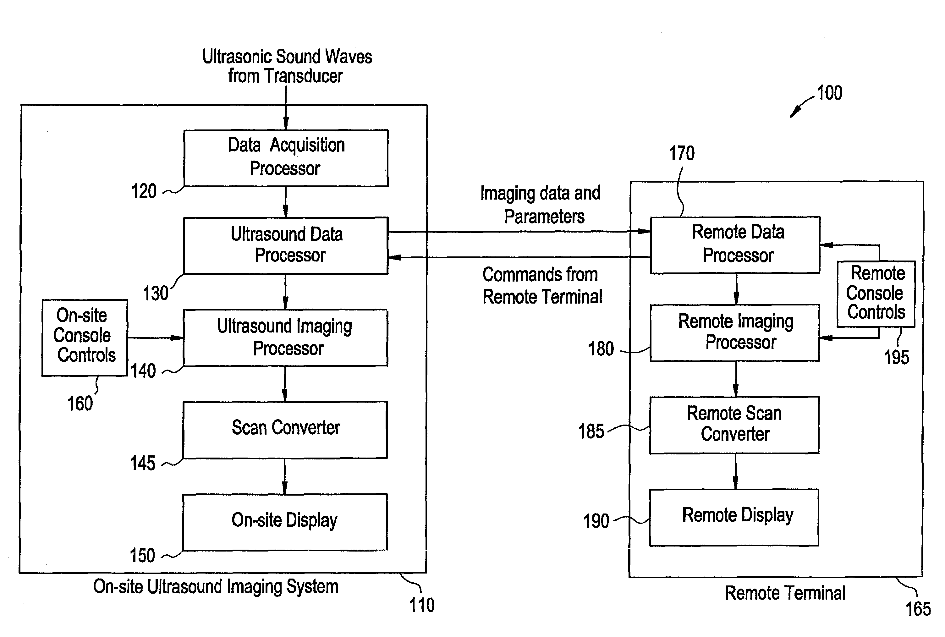
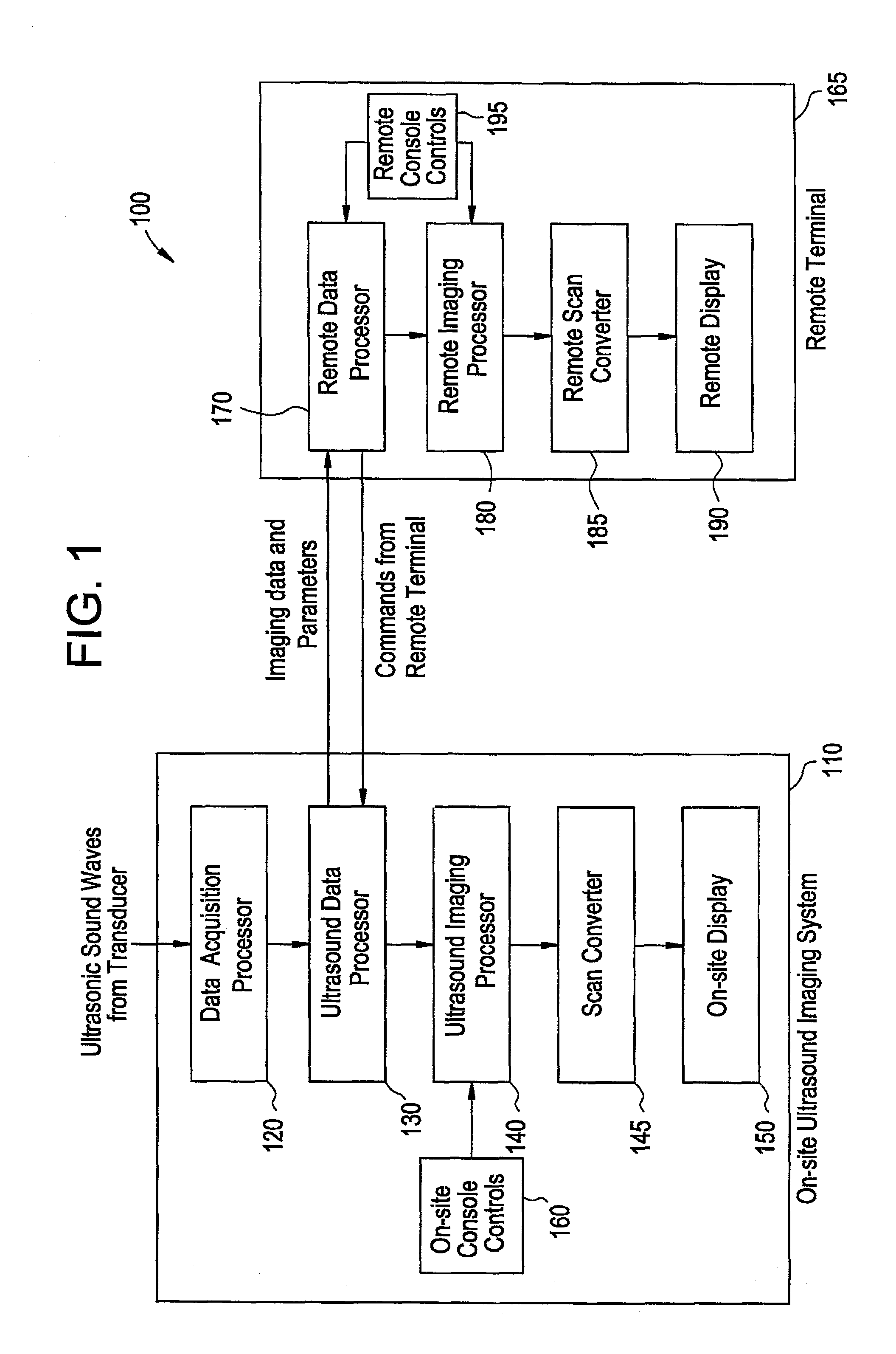
[0018]FIG. 1 illustrates a remotely controllable ultrasound imaging system 100 according to a preferred embodiment of the present invention. The imaging system 100 includes an ultrasound imaging system 110 and a remote terminal 165. The ultrasound imaging system includes a data acquisition processor 120, an ultrasound data processor 130, an ultrasound imaging processor 140, a scan converter 145, an on-site display 150, and on-site console controls 160. The remote terminal 165 includes a remote data processor 170, a remote imaging processor 180, a remote scan converter 185, a remote display 190, and remote console controls 195.

[0019]The ultrasound imaging system 110 of the remotely controllable ultrasound imaging system 100 is preferably attached to a transducer (not shown). The transducer is connected to an input port of the ultrasound imaging system 110. The data acquisition processor 120, the ultrasound data processor 130, the ultrasound imaging processor 140, and the scan convert...


PUM


Abstract

A system and method for streaming unprocessed medical image data from a medical imaging system to a remote terminal is provided. A medical imaging system acquires medical image data, generates unprocessed medical image data, and then transmits the unprocessed medical image data to a remote terminal. The remote terminal receives the unprocessed medical image data, processes the data to render a medical image and displays the medical image to an operator at the remote terminal. Additionally, the operator may control imaging parameters at the remoter terminal for use in rendering the medical image. Additionally, the operator may control imaging parameters on the medical imaging system. Also, the operator at the remoter terminal and the operator at the medical imaging system may communicate with each other during the examination through the medical imaging system.

Description

BACKGROUND OF THE INVENTION[0001]The present invention generally relates to a system and method for transmitting medical imaging data. More particularly, the present invention relates to transmitting remotely controllable unprocessed medical imaging data over a network such as the Internet, an Intranet, or a wireless network to a remote site for viewing by an expert.[0002]Medical imaging systems are typically used for a wide variety of applications in the field of medicine. For example, medical imaging systems such as ultrasound, CT scan, MRI, or X-ray systems, may be used for diagnosis or monitoring purposes. One type of medical imaging system commonly used in medicine is an ultrasound imaging system. Typical ultrasound imaging systems operate by transmitting ultrasonic sound waves into a patient's body using a transducer. The transducer is typically a device placed on the patient's body over the area to be imaged that is capable of sending and receiving ultrasonic sound waves. The...

Claims


Application Information

Patent Timeline
Patent Type & Authority Patents(United States)
IPC IPC(8): G06F15/16G06F19/00A61B5/00A61B5/055A61B6/00A61B8/00G01S15/89G06Q50/22H04N7/18
CPCA61B6/563A61B8/00A61B8/467A61B8/565G01S7/52084G01S15/899G06F19/321G06F19/3418G06Q50/22H04N1/32529H04N7/18A61B5/055A61B5/7232H04N2201/0075H04N2201/0081H04N2201/0089H04N2201/3225H04N2201/3278A61B6/00A61B8/461A61B8/468G16H30/20G16H40/67G16H30/40
Inventor KATSMAN, IGORSOKULIN, ALEXANDERKEMPINSKI, ARCADY
Owner GE MEDICAL SYST GLOBAL TECH CO LLC
Why Eureka
Try Eureka