Systems and methods for analyzing in vivo tissue volumes using medical imaging data

a tissue volume and imaging data technology, applied in the field of systems and methods for analyzing in vivo tissue volume using medical imaging data, can solve the problems of inability to meet the wide and variable clinical challenges of the current system or method, inability to optimally assess the disease through multi-site use, prohibitively expensive multi-disciplinary subspecialty assessment, etc., to facilitate clinical determination and/or pathological evaluation.

Inactive Publication Date: 2014-07-03
MAYO FOUND FOR MEDICAL EDUCATION & RES
View PDF1 Cites 29 Cited by
  • Summary
  • Abstract
  • Description
  • Claims
  • Application Information

AI Technical Summary

Problems solved by technology

In fact, one substantial challenge to the effective use of the wide and varied information available through non-invasive imaging is the ability to analyze, parse, and ultimate use particular pieces of the vast information provided in a given medical image to drive clinical decisions.
Similarly, an oft-cited survey paper on the “Computer Analysis of Computed Tomography Scans of the Lung” (IEEE TMI 25(4), April 2006: 385-405, states “First step toward more advanced processing schemes have been taken, but in the computer analysis of Diffuse Pulmonary Lung Disease, the question on what exactly to aim for and how to achieve it is still open.” The paper continues, “Classification and quantification of interstitial lung disease is difficult, and even experienced chest radiologists frequently struggle with different diagnoses.” However, “Automated schemes that indicate a percentage of affected lung or the probability of a certain disease would certainly be welcome, but require more research.” This portion of the paper concludes, “A quick analysis of the roughly 300 publications considered for this survey reveals that the amount of publications in this field has grown by a factor 1.5 per year over the past five years.” However, despite the proliferation of academic hype on the strategies for quantifying diseases such as lung diseases using medical images, none of the currently-available systems or methods is readily capable of meeting the wide and variable clinical challenges.
Such disturbing disparity could (at times, irreversibly) compromise patient care, and the optimal assessment of disease through the use of multi-site, multidisciplinary subspecialty assessment is prohibitively expensive and practically unfeasible.
Despite its efficacy, the consensus-based diagnosis has not attained clinical familiarity, let alone integration into routine practice.
Beyond the traditional barriers of physician adherence to clinical practice guidelines, the ATS / ERS recommendation lacks practicality.
It is impractical in typical clinical, or even in multispecialty academic settings, to routinely establish consensus via group discussion among multiple physicians.
Even if this was possible, the differences in experience, knowledge and potential unblinded bias could adversely affect the accuracy and consistency of such a consensus diagnosis.

Method used


Image

  • Systems and methods for analyzing in vivo tissue volumes using medical imaging data
  • Systems and methods for analyzing in vivo tissue volumes using medical imaging data
  • Systems and methods for analyzing in vivo tissue volumes using medical imaging data

Examples


Embodiment Construction

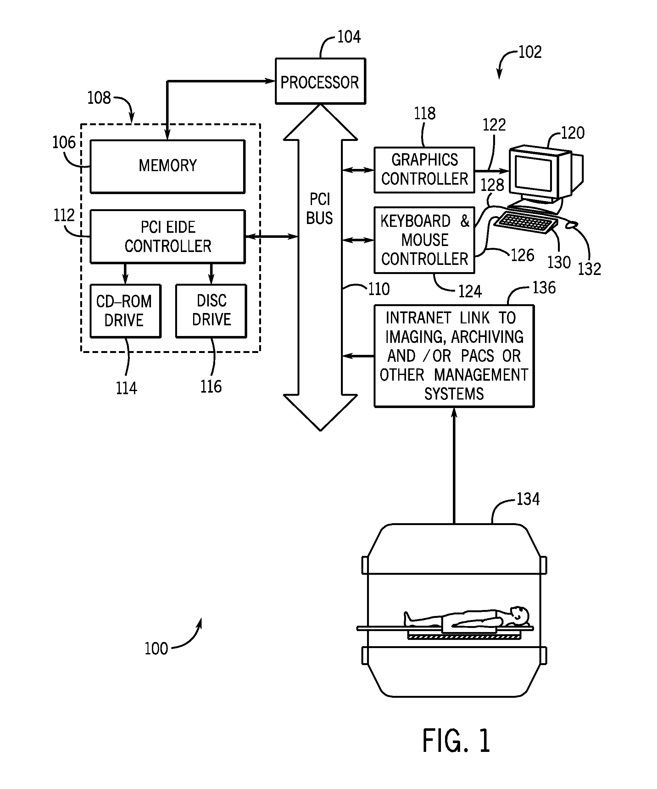
[0029]Referring now to FIG. 1, an analysis and imaging system 100 for conducting analysis in accordance with the present invention is illustrated. The system includes computer workstation 102 includes a processor 104 that executes program instructions stored in a memory 106 that forms part of a storage system 108. The processor 104 is a commercially available device designed to operate with available operating systems. It includes internal memory and I / O control to facilitate system integration and integral memory management circuitry for handling all external memory 106. The processor 104 also has access to a PCI bus driver that facilitates interfacing with a PCI bus 110.

[0030]The PCI bus 110 is an industry standard bus that transfers data between the processor 104 and a number of peripheral controller cards. These include a PCI EIDE controller 112 which provides a high-speed transfer of data to and from an optical drive 114 and a disc drive 116. A graphics controller 118 couples t...


PUM


Abstract

Computer-aided methods and computer-based systems designed to elicit information from imaging data of a volume of in vivo tissue to facilitate clinical determinations and / or pathological evaluation.

Description

CROSS-REFERENCE TO RELATED APPLICATIONS[0001]This application is based on, claims the priority to, and incorporates herein by reference U.S. Provisional Patent Application Ser. No. 61 / 518,424, entitled “SYSTEMS AND METHODS FOR ANALYZING IN VIVO TISSUE VOLUMES USING MEDICAL IMAGING DATA,” filed May 5, 2011, and U.S. Provisional Patent Application Ser. No. 61 / 483,881, entitled “SYSTEMS AND METHODS FOR ANALYZING IN VIVO TISSUE VOLUMES USING MEDICAL IMAGING DATA,” filed May 9, 2011.STATEMENT REGARDING FEDERALLY SPONSORED RESEARCH[0002]Not applicable.BACKGROUND OF THE INVENTION[0003]The present application is directed to systems and methods for analyzing in vivo tissue volumes using medical imaging data.[0004]Medical imaging has become a mainstay of modern clinical research and medicine. Medical images provide can provide a researcher or clinician with a wealth of information about in vivo anatomical structure and physiological performance and, thereby, provide key clinical indicators an...

Claims


Application Information

Patent Timeline
Patent Type & Authority Applications(United States)
IPC IPC(8): G06T11/20G06T7/00
CPCA61B5/055A61B6/032A61B6/037A61B6/5205A61B6/5217A61B5/7264A61B5/7267G06T11/206G06T7/0081G06T7/602G06T2207/10072G06T2207/30061G06T2207/30101G06T7/0012G06T7/11G06T7/62G16H50/30
Inventor ROBB, RICHARD A.RAJAGOPALAN, SRINIVASANKARWOSKI, RONALD A.BARTHOLMAI, BRIAN J.
Owner MAYO FOUND FOR MEDICAL EDUCATION & RES
Why Eureka
Try Eureka