Ultrasound diagnostic apparatus and method

A diagnostic device and ultrasonic technology, applied in the directions of sonic diagnosis, infrasonic diagnosis, ultrasonic/sonic/infrasonic diagnosis, etc., can solve the problems of difficult compression, effective imaging object, difficult elastic rate, etc., and achieve the effect of high safety

Inactive Publication Date: 2013-10-09
HITACHI LTD
View PDF12 Cites 23 Cited by
  • Summary
  • Abstract
  • Description
  • Claims
  • Application Information

AI Technical Summary

Problems solved by technology

In this method, as the imaging target, although it is extremely effective for organs (such as breasts, etc.) that exist in parts that are relatively easy to be compressed from the body surface, it may not be effective for all imaging targets.
For example, as an intermediate layer between the body surface and the liver, it is difficult to generate sufficient displacement such compression due to the presence of sliding surfaces
In addition, in the conventional method, when it is desired to estimate the elastic rate, boundary conditions are necessary, but if there is a sliding surface, the boundary conditions become complicated and it is difficult to estimate the elastic rate

Method used


Image

  • Ultrasound diagnostic apparatus and method
  • Ultrasound diagnostic apparatus and method
  • Ultrasound diagnostic apparatus and method

Examples


Embodiment 1

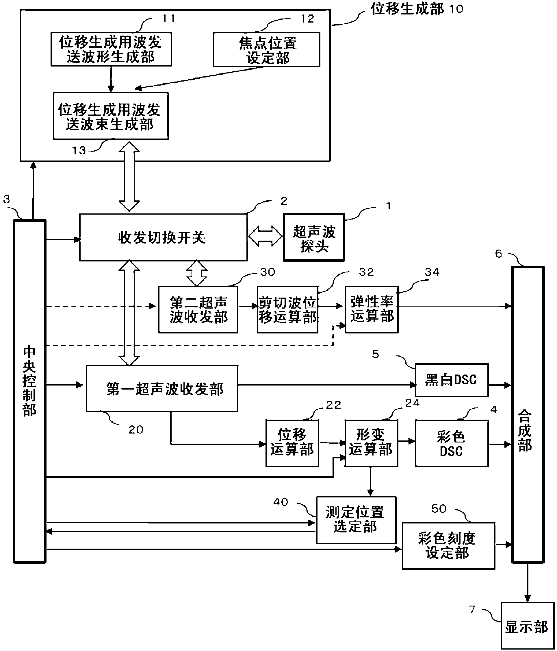
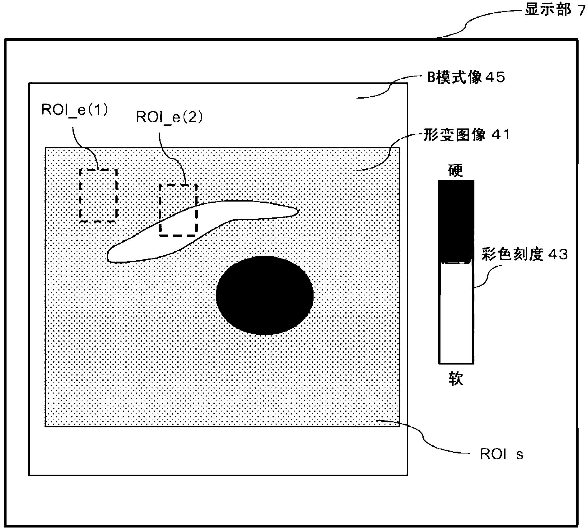
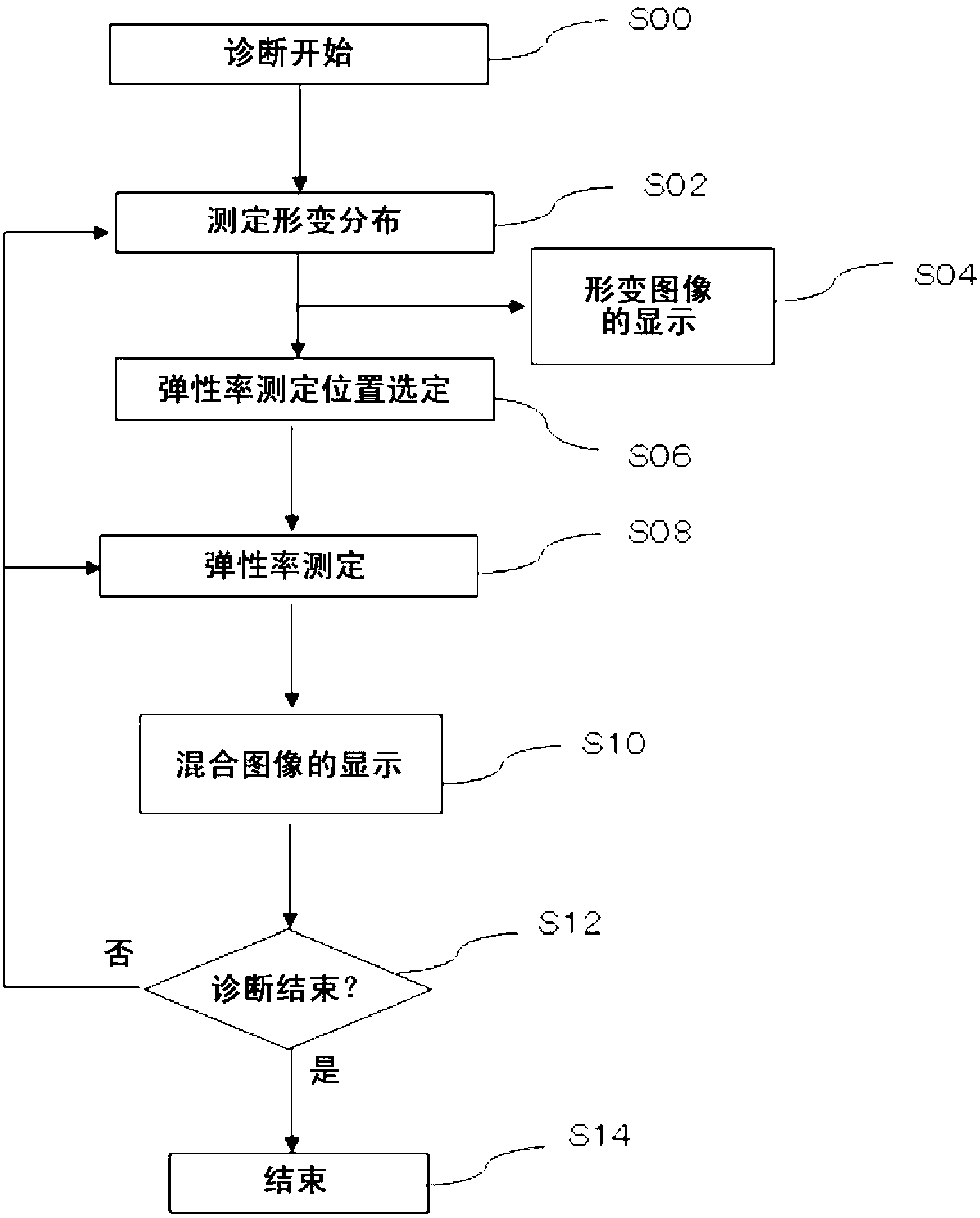
[0045] Embodiment 1 relates to an ultrasonic diagnostic apparatus that uses an ultrasonic probe that transmits an ultrasonic beam to a subject and receives an echo signal, and includes: a deformation calculation unit 24 that radiates a first displacement detection beam and based on Calculate the deformation information in the region 1 from the echo signal of the subject subjected to wave reception; the displacement generation unit 10 irradiates the focused beam into the subject to displace the tissue in the subject; the elastic rate calculation unit 34, It radiates the second displacement detection beam, detects the displacement of the shear wave generated by the focused beam based on the echo signal from the subject subjected to wave reception, and detects the displacement of the shear wave included in the region 1. elastic modulus; and a display section 7 that displays a deformation image and elastic modulus based on the deformation information.

[0046] In addition, the pre...

Embodiment 2

[0101] Embodiment 2 relates to an ultrasonic diagnostic apparatus that uses an ultrasonic probe that transmits an ultrasonic beam to an object and receives an echo signal. a beam for calculating deformation information in the region 1 based on an echo signal from the subject subjected to wave reception; a displacement generation unit for radiating the focused beam into the subject to cause displacement of the tissue in the subject; elastic modulus The calculation unit 34 radiates the second displacement detection beam, detects the displacement of the shear wave generated by the focused beam based on the echo signal from the subject subjected to wave reception, and detects the displacement of the shear wave included in the region 1 The elastic modulus in the area 2; the display unit 7, which displays the deformation image and the elastic modulus based on the deformation information; and the measurement position selection unit 40, which detects the elastic modulus based on the de...

Embodiment 3

[0152] Next, as a third example, a hybrid system in which example 1 and example 2 are combined will be described. The system configuration of the third embodiment not shown is in figure 1 Added to the system configuration Figure 10 The beam time setting unit 14 and the hardness spectrum calculation unit 35 are formed. The output signal from the shear wave displacement calculation unit 32 is input to both the elastic modulus calculation unit 34 and the hardness spectrum calculation unit 35 which are provided in parallel. In addition, the central control unit 3 is connected to both the beam time setting unit 14 and the hardness spectrum calculating unit 35 . The operator selects any one of the modulus of elasticity calculation unit 34 and the hardness spectrum calculation unit 35 arranged in parallel. In this specification, the modulus of elasticity calculating unit 34 and the hardness spectrum calculating unit 35 may be collectively referred to as an modulus of elasticity c...


PUM


Abstract

Provided is an ultrasound diagnostic apparatus with which elastic modulus can be safely measured with high accuracy over a wide area when measuring elastic modulus using shear wave generation. The ultrasound diagnostic apparatus comprises: an ultrasound probe (1) that sends and receives echo signals from inside a living body; a strain-computing unit (24) that receives an echo signal from the body by radiating a first displacement-detecting beam and computes strain information in a Region 1; a measurement position-selecting unit (40) that selects a Region 2, which is contained within Region 1 and is smaller than Region 1, as the elastic modulus-measurement position on the basis of the strain information; a displacement-generating unit (10) that radiates a focused beam that is the displacement-generating transmission beam into the body and displaces the tissue; an elastic modulus-computing unit (34) that receives an echo signal from the body by radiating a second displacement-detecting beam, detects the shear wave displacement that results from the focused beam, and computes the elastic modulus in Region 2; and a display unit (7) that displays the strain image that is based on the strain information in Region 1 and the elastic modulus. The display unit (7) also displays a color scale that represents the elastic modulus in the display area of the displayed strain image.

Description

[0001] technology area [0002] The present invention relates to an ultrasonic diagnostic apparatus for diagnosing a subject by transmitting and receiving ultrasonic waves, and more particularly to an ultrasonic diagnostic technique for detecting differences in hardness inside a subject. Background technique [0003] As a method for diagnosing breast cancer, liver cirrhosis, and vascular diseases in a living body, there is a method of diagnosing the hardness (deformation, elastic coefficient, etc.) method (elastography: elastography). In the hardness diagnosis based on the elastography technique, the researcher pushes the ultrasonic probe to the surface of the subject and compresses it to displace the tissue inside the living body (hereinafter, this method will be referred to as a conventional method). Estimate the displacement in the compression direction based on the echo signals of the biological tissue before and after compression caused by compression, calculate the spat...

Claims


Application Information

Patent Timeline
Patent Type & Authority Applications(China)
IPC IPC(8): A61B8/08
CPCA61B8/42A61B8/469A61B8/485A61B8/5246G01S7/52042
Inventor 田原麻梨江东隆吉川秀树桥场邦夫
Owner HITACHI LTD
Why Eureka
Try Eureka