
Cervical vertebra flexed position magnetic resonance imaging fixing device

A fixed device, magnetic resonance technology, applied in medical science, sensors, diagnostic recording/measurement, etc., can solve the problems of reducing the quality of image diagnosis, non-standard scanning imaging of patients' flexion position, etc., achieving novel structure, convenient scanning, and automatic adjustment. Effect of head and neck flexion angle

Inactive Publication Date: 2015-03-11
侯红军
View PDF6 Cites 3 Cited by
  • Summary
  • Abstract
  • Description
  • Claims
  • Application Information

AI Technical Summary

Problems solved by technology

Therefore, MRI in the flexion position of the cervical spine is of great value in the early diagnosis of Hirayama disease, the exploration of its pathogenesis, and the follow-up after treatment. For clinical diagnosis, the patient is usually placed on the flat pad of the cervical spine scanning coil placed on the scanning table for magnetic resonance imaging. Obtain cervical spine imaging in a natural position to meet disease diagnosis requirements. This cervical spine scan coil flat pad is composed of a head and neck pad, a chest and back pad, and a scan coil lumbosacral pad. The two sides of the head and neck pad are respectively fixed with a cervical spine scan coil. The disadvantage of this structure is that when MRI examination in the flexion position of the cervical spine is required, non-metallic objects can only be placed under the top of the patient's head. Due to the randomness of the objects placed under the top of the head, there will be scans of the patient in flexion position. Not standard, at the same time, due to the uncomfortable shaking of the head and neck of the patient during the scanning imaging process, scanning motion artifacts appear, which greatly reduces the quality of image diagnosis

Method used


Image

  • Cervical vertebra flexed position magnetic resonance imaging fixing device
  • Cervical vertebra flexed position magnetic resonance imaging fixing device
  • Cervical vertebra flexed position magnetic resonance imaging fixing device

Examples


Embodiment Construction

[0018] Below in conjunction with accompanying drawing, the present invention is further described:

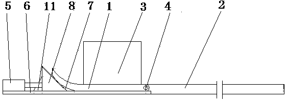
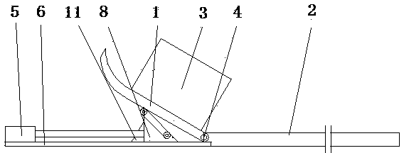
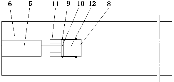
[0019] As shown in the accompanying drawings, a cervical spine flexion magnetic resonance examination fixation device includes a head and neck pad 1, a chest and back pad 2, and a backing board 3, and a magnetic resonance scanner is embedded in the head and neck pad 1, the chest and back pad 2, and the backing board 3 The coil is characterized in that it is provided with a rotating shaft 4, a telescopic cylinder 5, a base plate 6 and a buckling support device 7, the front end of the head and neck pad 1 is hinged to the chest and back pad 2 through the rotating shaft 4, and scanning coil backing boards 3 are respectively provided on both sides, The lower part of the head and neck pad 1 is provided with a base plate 6, and the base plate 6 is provided with a buckling support device 7. One end of the base plate 6 is fixedly connected with the chest and back pad 2, and the other end...


PUM


Abstract

The invention discloses a cervical vertebra flexed position magnetic resonance imaging fixing device. The device comprises a head-neck cushion, a chest-back cushion and backup plates, wherein magnetic resonance imaging coils are respectively embedded in the neck cushion, the chest-back cushion and the backup plates. The device is characterized by comprising a rotating shaft, a telescopic air cylinder, a bottom plate and a flexion supporting device; the front end of the head-neck cushion is hinged to the chest-back cushion through the rotating shaft; the scanning coil backup plates are respectively arranged on two sides of the head-neck cushion; the bottom plate is arranged at the lower part of the neck cushion; the flexion supporting device is arranged on the bottom plate; one end of the bottom plate is connected with the chest-back cushion, and the other end of the bottom plate is fixedly connected with the telescopic air cylinder; a guide slide way is arranged in the center of the bottom plate; the flexion supporting device comprises a supporting wedge block, a rolling shaft and a supporting foot; the rolling shaft is arranged on the inclined front surface of the supporting wedge block; a guide rail is arranged on the lower bottom surface of the supporting wedge block; a support roller is arranged on the rolling shaft; the inclined rear surface of the supporting wedge block is fixedly connected with the telescopic air cylinder; the inclined front surface of the supporting wedge block is in contact with the head-neck cushion; the lower bottom surface of the supporting wedge block is connected with the guide slide way. The device has the advantages of novel structure, convenience in scanning, high image quality and the like.

Description

technical field [0001] The invention relates to the technical field of medical equipment, in particular to a cervical spine flexion magnetic resonance examination fixation device for assisting magnetic resonance equipment examination. Background technique [0002] Hirayama disease is also called distal upper extremity muscular atrophy in youth. It is a benign self-limited motor neuron disease first reported by Japanese scholar Keizo Hirayama in 1959. It is clinically similar to motor neuron disease amyotrophic lateral sclerosis and progressive muscular atrophy of the spinal cord, but its prognosis is completely different. Primary disease is prone to occur in adolescence, and male is more common. The ratio of male to female is about 20:1. Muscle atrophy refers to rhabdomyotrophy, the muscle volume is smaller than normal, and the muscle fibers become thinner or even disappear. Neuromuscular hypertrophy. In addition to the pathological changes of muscle tissue itself, musc...

Claims


Application Information

Patent Timeline
Patent Type & Authority Patents(China)
IPC IPC(8): A61B5/055
Inventor 侯红军许东
Owner 侯红军
Learn More