A Segmentation Method for Inhomogeneous Medical Images

A medical image and uniform technology, applied in the field of image analysis, can solve problems such as unrealizable segmentation and unrealizable segmentation, and achieve the effect of reducing dependence, realizing accurate segmentation, and precise segmentation

Inactive Publication Date: 2017-03-08
SOUTHERN MEDICAL UNIVERSITY
View PDF2 Cites 2 Cited by
  • Summary
  • Abstract
  • Description
  • Claims
  • Application Information

AI Technical Summary

Problems solved by technology

However, this method only uses the homogeneous area as a node, and the generation of auxiliary seed points also depends on the homogeneous area. Therefore, when the target to be segmented is heterogeneous, the non-homogeneous area of ​​the target area cannot be well segmented. Still can't achieve accurate segmentation

Method used


Image

  • A Segmentation Method for Inhomogeneous Medical Images
  • A Segmentation Method for Inhomogeneous Medical Images
  • A Segmentation Method for Inhomogeneous Medical Images

Examples


example 1

[0039] Example 1 (Segmentation of non-uniform two-dimensional medical images)

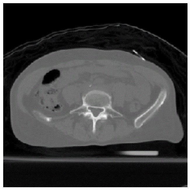
[0040] This embodiment is based on figure 1 A CT (Computed Tomography) image of the abdomen of a patient is shown as an example to describe the implementation process of the method of the present invention. figure 1 The size of the lumbar vertebrae is 512×512, and the lumbar vertebrae to be segmented belong to the non-uniform target, which includes high-density cortical bone, medium-density bone compact and low-density bone marrow. The specific segmentation method is as follows:

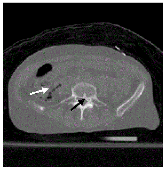
[0041] Step 1: Read in as figure 1 For the CT image shown, through the MATLAB GUI graphical interface, select the foreground seed points that can represent the lumbar vertebrae and the background seed points that represent other normal tissues of the abdomen. Superimpose the selected seed points on figure 1 On, get as figure 2 In the illustrated abdomen CT image of the lumbar vertebrae to be segmented in the marked seed point...

example 2

[0050] Example 2 (Segmentation of non-uniform 3D medical images)

[0051] This embodiment is based on Figure 8 The illustrated pelvic CT image containing the applicator taken when a certain cervical cancer patient undergoes brachytherapy afterloading radiotherapy is taken as an example to describe the method of the present invention for the segmentation process of a non-uniform three-dimensional medical image. Figure 8 The size is 256×256×55. The applicator in the picture is a non-uniform three-dimensional target, which includes high-density metal pipes, medium-density plastics, and low-density liquids and air. The specific segmentation method is as follows:

[0052] Step 1: Read in as Figure 8 In the CT image of the pelvis shown, through the MATLAB GUI graphical interface, select the foreground seed point representing the applicator and the background seed point representing other tissue structures of the pelvis. Superimpose the selected seed points on Figure 8 On, get as Pic...


PUM


Abstract

The invention relates to a method for segmenting an inhomogeneous medical image. The method for segmenting the inhomogeneous medical image comprises the following steps that firstly, foreground seed points and background seed points on the image to be segmented are selected; secondly, the probability that each grey level belongs to the foreground or the background of the image to be segmented is evaluated according to grey level information of a selected seed point set, the grey levels are mapped on all pixel points of the image, and therefore a corresponding probability density distribution graph is obtained; thirdly, the selected foreground seed points and the selected background seed points are used as growing seed points respectively, one probability threshold on the corresponding probability density distribution graph is used as a growing condition, a region growing algorithm is executed, and therefore a foreground seed point group and a background seed point group which have grown automatically are obtained; finally, the obtained seed point groups which have grown automatically are used as seed points of a random walk algorithm, the random walk algorithm is executed, and a final segmentation result is obtained. By the adoption of the method for segmenting the inhomogeneous medical image, the sensitivity to the number and the position of initial seed points can be reduced, and the segmentation precision of the inhomogeneous medical image is obviously improved.

Description

Technical field [0001] The present invention relates to image analysis, in particular to a segmentation method of medical images. Background technique [0002] With the rapid development of imaging medicine, the segmentation of medical images is of great significance for clinical diagnosis and treatment. The current image segmentation algorithms are mainly divided into three categories: manual segmentation, interactive segmentation and automatic segmentation. Manual segmentation is time-consuming and requires the segmenter to have rich experience. Fully automatic segmentation does not require manual intervention, and is generally more suitable for the segmentation of simple and uniform images. However, for the segmentation of complex and diverse medical images, the accuracy of segmentation usually does not meet clinical needs. The interactive segmentation only needs to add a small amount of manual intervention to automatically segment a better result. In order to meet the segm...

Claims


Application Information

Patent Timeline
Patent Type & Authority Patents(China)
IPC IPC(8): G06T7/10G06T7/136G06T7/194
Inventor 陈海斌周凌宏甄鑫王琳婧肖阳胡洁
Owner SOUTHERN MEDICAL UNIVERSITY
Why Eureka
Try Eureka