Microendoscopic soft tissue suture guiding device and microendoscopic soft tissue suture guiding method

A guiding device and soft tissue technology, applied in medical science, surgery, surgical instruments, etc., can solve the problems of difficult puncture location positioning, soft tissue damage, prolonged operation time, etc., to avoid blind needle sticking, easy operation, and clear vision.

Active Publication Date: 2014-06-18
AFFILIATED HUSN HOSPITAL OF FUDAN UNIV
View PDF6 Cites 2 Cited by
  • Summary
  • Abstract
  • Description
  • Claims
  • Application Information

AI Technical Summary

Problems solved by technology

The two-dimensional image displayed by the endoscope cannot fully display the three-dimensional space of the joint capsule, and there are often multiple blind spots, which increases the difficulty of soft tissue repair
Due to the difficulty in locating the puncture position, the soft tissue is often damaged again during the tentative needling, which affects the healing ability of the soft tissue. Repeated attempts will prolong the operation time and increase the pain of the patient, making it difficult for various high-tech operations to be carried out in primary hospitals.

Method used


Image

  • Microendoscopic soft tissue suture guiding device and microendoscopic soft tissue suture guiding method
  • Microendoscopic soft tissue suture guiding device and microendoscopic soft tissue suture guiding method
  • Microendoscopic soft tissue suture guiding device and microendoscopic soft tissue suture guiding method

Examples


Embodiment 1

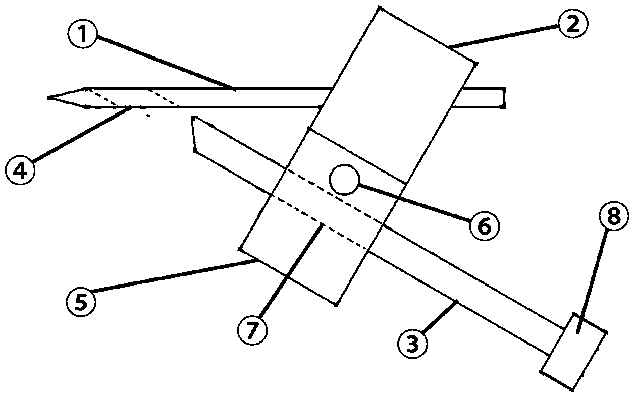
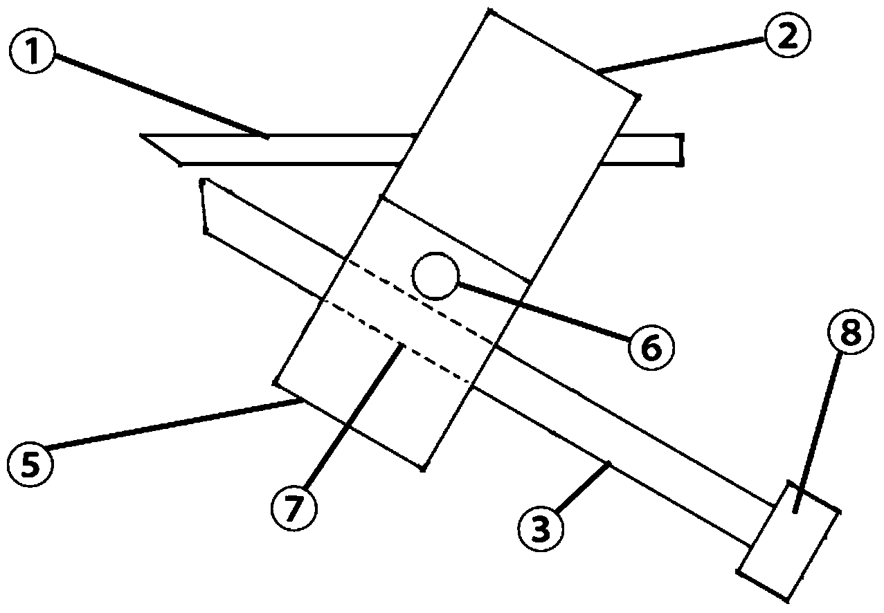
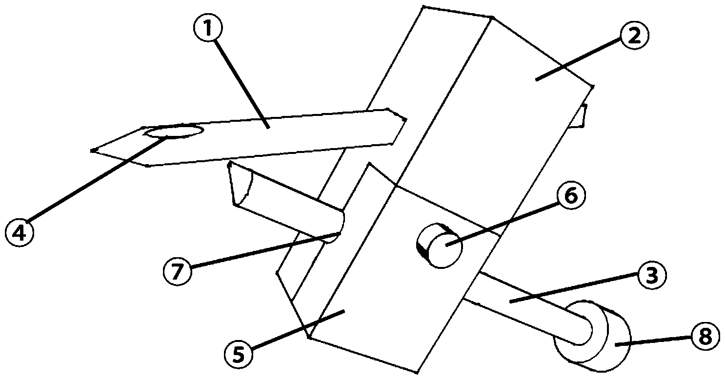
[0069] Such as Figure 12-13 As shown, there is a tear on the side of the soft tissue 11 close to the capsule wall 10, and the needle tip of the guide needle 1 is pierced into the joint capsule through the skin 9 and the capsule wall 10, pushed to the top of the planar soft tissue 11, and the needle tip of the guide needle 1 is inserted into the joint capsule. Place it at the target needle exit point of the soft tissue 11; after the needle tip of the hollow needle 3 pierces the skin 9 and the capsule wall 10, insert the part of the hollow needle 3 exposed outside the skin into the upper guide tube 7 of the guide body 2, and then remove the The block 5 is assembled and fixed with the guide body 2 to form a complete guide tube 7, which plays the role of clamping and restraining the hollow needle 3 and guiding it to the soft tissue 11; after the hollow needle 3 is installed into the complete guide tube 7, the hollow needle 3 push the needle tip to the target needle exit point of ...


PUM


Abstract

The invention relates to a microendoscopic soft tissue suture guiding device and a microendoscopic soft tissue suture guiding method. The microendoscopic soft tissue suture guiding device comprises a guide pin, a guide body and a hollow pin, wherein one end of the guide pin is provided with a pinpoint, and the other end of the guide pin is fixedly arranged in one end of the guide body; an included angle which is smaller than 90 degrees is formed between the other end of the guide pin and the guide body; the middle of the guide body is provided with a guide pipe; the hollow pin penetrates through the interior of the guide pipe; one end of the hollow pin is provided with a push handle, and a pinpoint at the other end of the hollow pin penetrates out of the pinpoint of the guide pin along the guide pipe. The device and the method can be used for simplifying the microendoscopic suture operation performed for treating soft tissue lacerated wounds; when puncturation or pin insertion is performed upward from an endoscope blind zone below a soft tissue, the device can be used for accurately locating a puncturation position, avoiding secondary soft tissue damages caused when trial pin insertion is performed as the puncturation position is difficult to locate; the device is simple in structure, convenient to operate, economical and practical, and convenient to popularize and apply.

Description

technical field [0001] The invention belongs to the technical field of medical soft tissue repair, in particular to an endoscopic soft tissue suturing guide device and method. Background technique [0002] Soft tissue laceration is a common disease in sports injuries, such as meniscus tear of knee joint and triangular fibrocartilage complex (TFCC) tear of wrist joint. With the development and updating of endoscopic equipment, the repair of soft tissue has been transitioned from open surgery to minimally invasive surgery. The two-dimensional image displayed by the endoscope cannot fully display the three-dimensional space of the joint capsule, and there are often multiple blind spots, which increases the difficulty of soft tissue repair. Due to the difficulty in locating the puncture site, tentative acupuncture often causes soft tissue damage again, affecting the healing ability of soft tissue. Repeated attempts will prolong the operation time and increase the pain of patien...

Claims


Application Information

Patent Timeline
Patent Type & Authority Applications(China)
IPC IPC(8): A61B17/04
CPCA61B17/00234A61B17/0469A61B17/0482
Inventor 徐文东尹华伟蒋苏沈云东徐建光顾玉东
Owner AFFILIATED HUSN HOSPITAL OF FUDAN UNIV
Why Eureka
Try Eureka