Structure and use of Japanese encephalitis virus envelope protein binding peptide
A technology of Japanese encephalitis virus and envelope protein, which is applied in the field of biomedicine to achieve good brain tissue targeting, important social and economic benefits, and increased uptake rate
- Summary
- Abstract
- Description
- Claims
- Application Information
AI Technical Summary
Problems solved by technology
Method used
Image
Examples
Embodiment 1
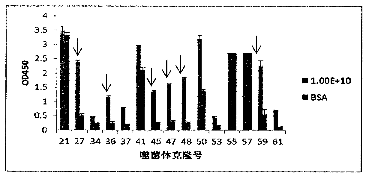
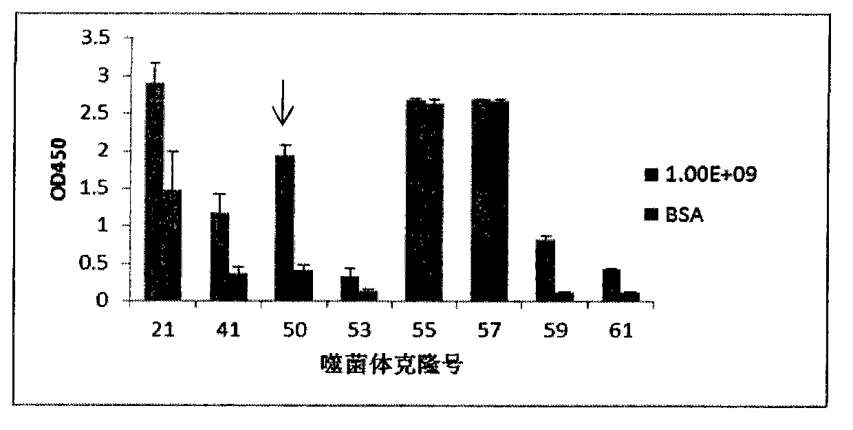
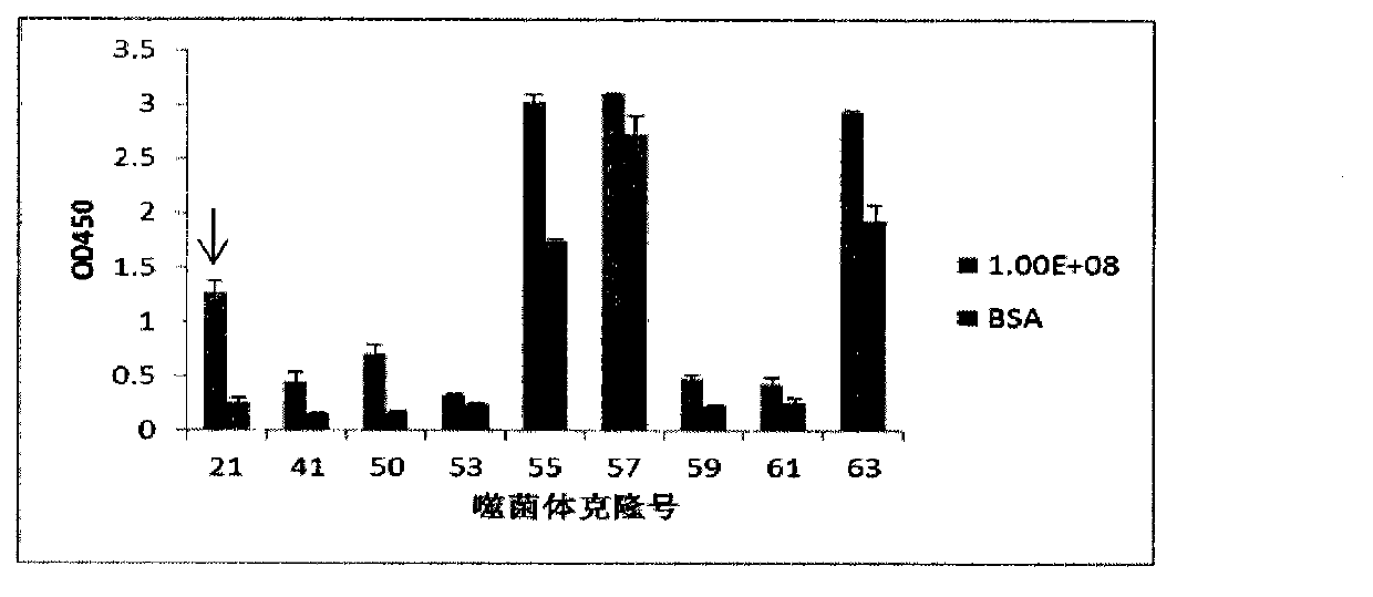
[0039] This example mainly illustrates the screening of envelope protein-binding polypeptides.
[0040] Materials and Methods
[0041] 1. Phage Random Peptide Library Screening
[0042] Using NaHCO 3 (PH=8.6) solution Coat 100 μl of 100 μg / mL envelope protein on a 96-well ELISA plate, and place in a humid environment at 4° C. overnight. The next day, the coating solution was poured off, and 300 μL of BSA blocking solution with a concentration of 10 mg / mL was added to each well, and blocked at 37°C for 1 hour. Wash 6 times with TBST (TBS+0.5% (v / v) Tween-20). Join 1.5×10 11 pfu (in 100 μL TBST) phage (Ph.D.-12 TM Phage Display Peptide Library Kit (purchased from New England Biolabs), combined at 37°C for 1 hour, decanted the phage, and washed 10 times with TBST (TBS+0.5% (v / v) Tween-20). Use 100μl of suitable eluent (0.2M Glycine-HCl (pH2.2), 1mg / ml BSA) to elute for 7-9min at room temperature, not more than 10min, suck out the eluent into a centrifuge tube, add 15μL of ...
Embodiment 2
[0048] This example mainly illustrates the uptake of 11 binding peptides in virus-infected and uninfected cells.
[0049] Materials and Methods
[0050] 1. BHK21 cells, viruses, polypeptides JE virus is amplified by infecting BHK21 cells, then mixed, subpackaged and stored at -70°C for later use. The JE virus strain used was Jingweiyan strain. BHK21 cell culture medium is DMEM (GIBCO) medium containing 7% fetal bovine serum. A maintenance solution containing 0.7% fetal bovine serum was used for virus infection. 11 peptides were labeled with FITC during synthesis.
[0051] 2. Quantitative observation of polypeptide uptake rate in virus-infected and uninfected cells by flow cytometry BHK21 cells were cultured in DMEM medium (GIBCO) containing 7% fetal bovine serum at 37°C and 5% CO2 incubator. It was observed that the cells were in good growth state. After culturing to the logarithmic growth phase, the cells were inoculated into a 6-well cell culture plate, and 80-90% of the...
Embodiment 3
[0055] This example mainly illustrates the uptake rates of 11 binding peptides in the brains of virus-infected and uninfected mice.
[0056] Materials and Methods
[0057] Order a number of SPF three-week-old (9-11g) BALB / C female mice, weigh and group the mice in advance at the beginning of the experiment, and prepare the experimental things: each FITC-labeled polypeptide, 1mL syringe, cotton swab, electronic Name, record book. The polypeptide group was injected intraperitoneally at a dose of 20 mg / kg, and the mice in the polypeptide virus group were intraperitoneally injected with a dose of 20 mg / kg immediately after challenge. The mice were sacrificed 4 hours later, and the brain tissue was immediately taken out, fixed in 4% formaldehyde, and sent for in vivo imaging to observe the fluorescence intensity of the brain tissue.
[0058] result
[0059] In vivo imaging results showed that peptides P63, P45, P55, P36, P50, and P48 have good targeting properties in virus-infec...
PUM
 Abstract
Description
Claims
Application Information
 - R&D Engineer
- R&D Manager
- IP Professional
- Industry Leading Data Capabilities
- Powerful AI technology
- Patent DNA Extraction
Browse by: Latest US Patents, China's latest patents, Technical Efficacy Thesaurus, Application Domain, Technology Topic.
© 2024 PatSnap. All rights reserved.Legal|Privacy policy|Modern Slavery Act Transparency Statement|Sitemap


