A method for establishing goose embryo epithelial cell line and established goose embryo epithelial cell line

A technology of epithelial cells and goose embryos is applied in the field of established goose embryo epithelial cell lines to achieve the effect of less restriction, uniform shape and high purity

Active Publication Date: 2019-05-17
SHANDONG BINZHOU ANIMAL SCI & VETERINARY MEDICINE ACADEMY
View PDF2 Cites 1 Cited by
  • Summary
  • Abstract
  • Description
  • Claims
  • Application Information

AI Technical Summary

Problems solved by technology

However, there is no commercially available goose-derived cell line

Method used


Image

  • A method for establishing goose embryo epithelial cell line and established goose embryo epithelial cell line
  • A method for establishing goose embryo epithelial cell line and established goose embryo epithelial cell line
  • A method for establishing goose embryo epithelial cell line and established goose embryo epithelial cell line

Examples


Embodiment 1

[0078] The establishment of embodiment 1 goose embryo epithelial cell line

[0079] Establish the goose embryo epithelial cell line of the present invention according to the following steps:

[0080] (1) Aseptically collect 11-13-day-old goose embryo tissues, cut off the head, limbs and internal organs with sterilized scissors, put them into a 10-mL glass beaker, add PBS at a concentration of 10 mL, and wash for 5-10 minutes; discard After PBS, add 10-20 mL of gentamicin solution with a mass percentage concentration of 20%-70% prepared in PBS and treat for 5-10 minutes;

[0081] (2) Take out the duck embryo tissue and cut it into 0.5-1.5mm3 tissue pieces, put the tissue pieces flat into the bottom of the wells of the 6-well culture plate, one piece for each well, add 2-3mL containing 10% fetal bovine by volume to the culture wells Serum, 1%-2% goose serum and DMEM culture medium of 0.2%-1% goose embryo allantoic fluid, cultured at 37°C and 5% CO2 until the tissue pieces are c...

Embodiment 2

[0092] The biological characteristic analysis of embodiment 2 goose embryo epithelial cell lines

[0093] 1. Morphological observation

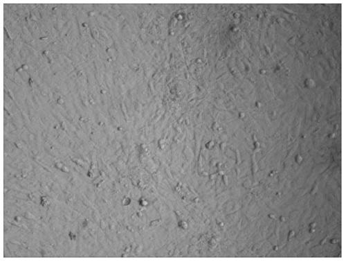
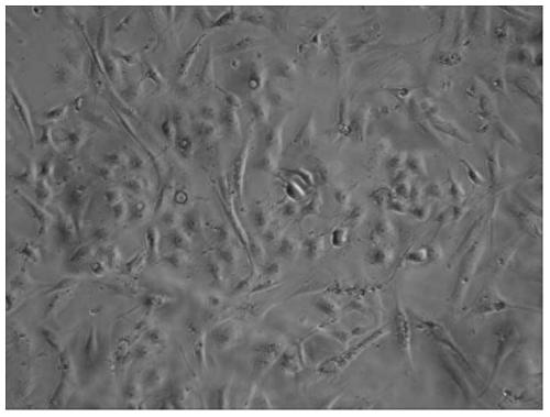
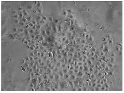
[0094] Observed by an inverted microscope, the established goose embryo epithelial cell line F30 generation ( Figure 4 ) and the F50 generation ( Figure 5 ) and primary cells ( figure 1 ) compared with the morphological difference, the primary cells contain a variety of miscellaneous cells, most of the cells are long fusiform, a few are polygonal and oval, and the established epithelial cell line is obtained from the monoclonal cell, and its purity Higher than 99.9%, all are epithelioid cells, and HE staining also shows that the cells are polygonal or short spindle-shaped epithelioid cells ( Figure 6 ), with round nuclei, strong cell growth and division abilities, and a population doubling time of only 17.1 hours.

[0095] 2. Growth curve determination

[0096] Take the 30th and 50th generation of cells in good growth state, and when ...

Embodiment 3

[0102] Example 3 Sensitivity Verification of Goose Embryo Epithelial Cell Lines to Muscovy Duck Parvovirus

[0103] 1. Method

[0104] Take the 50th generation cells of the goose embryo epithelial cell line, discard the culture medium when it grows into 80% monolayer, wash it twice with D-Hanks medium, inoculate Muscovy duck parvovirus, absorb at 37°C for 1 hour, and discard the virus liquid And supplemented with DMEM maintenance solution (containing 1% newborn bovine serum) for culture, and a blank control (goose embryo epithelial cell line not inoculated with Muscovy duck parvovirus) was set at the same time. The same method was used to inoculate goose plague virus, type I duck hepatitis virus, and new duck hepatitis virus.

[0105] 2. Results

[0106] The epithelial cells of the goose embryos were observed to be sensitive to the Muscovy duck parvovirus, and the infected cells all produced specific cytopathic changes within 96 hours, see Figure 11 ; while the control cel...


PUM


Abstract

The invention discloses a method for establishing goose embryo epithelial cell line and the established goose embryo epithelial cell line. The invention relates to the method for establishing the goose embryo epithelial cell line, which is characterized in that a primary goose tissue adherent method, a differential velocity enzymatic digestion and a monoclonal screening method are combined, so that primary culture condition can be optimized. The method has the advantages of simple operation process, and convenient popularization and application. The invention also relates to the goose embryo epithelial cell line established by the method, a preservation number of the goose embryo epithelial cell line is CCTCC NO: C2014137, The establishment of the goose embryo epithelial cell line solves the problem of no well-established goose source cell line in prior art. The invention also relates to a kit used for culturing and / or proliferating goose parvovirus, muscovy duck parvovirus and type I duck hepatitis virus and novel duck hepatitis virus, and is characterized in that a host cell to be infected is / or comprises the goose embryo epithelial cell line. The method for establishing the goose embryo epithelial cell line verifies the sensitive characteristic of the goose parvovirus, muscovy duck parvovirus and type I duck hepatitis virus and novel duck hepatitis virus.

Description

technical field [0001] The invention belongs to the technical field of animal cell engineering, and in particular relates to a method for establishing a goose embryo epithelial cell line and the established goose embryo epithelial cell line. Background technique [0002] my country is a big country of waterfowl production, and waterfowl output accounts for more than 60% of the world, wherein the output of geese accounts for more than 90% of the world's total output. my country has a long history of raising geese. According to textual research, geese were domesticated as early as the Neolithic Age about 6,000 years ago. With the development of my country's economy and the continuous improvement of people's living standards, the increase in the proportion of animal food consumption and the improvement of consumption habits have promoted the adjustment of the agricultural industrial structure and the improvement of the production status of animal husbandry. , Less food consumpt...

Claims


Application Information

Patent Timeline
Patent Type & Authority Patents(China)
IPC IPC(8): C12N5/073C12N7/00C12R1/91
Inventor 王文秀沈志强于金枝
Owner SHANDONG BINZHOU ANIMAL SCI & VETERINARY MEDICINE ACADEMY
Why Eureka
Try Eureka