Method of determining the presence or absence of target nucleic acid in cell sample

A target nucleic acid and cell technology, applied in biochemical equipment and methods, microbial measurement/inspection, DNA preparation, etc., can solve problems such as extending running time

Inactive Publication Date: 2015-04-29
QIAGEN GMBH
View PDF16 Cites 3 Cited by
  • Summary
  • Abstract
  • Description
  • Claims
  • Application Information

AI Technical Summary

Problems solved by technology

This extended run time prevented the customer from processing a full tray of 88 samples plus calibrators and controls, including hc assays, in one shift (8 hours)

Method used


Image

  • Method of determining the presence or absence of target nucleic acid in cell sample
  • Method of determining the presence or absence of target nucleic acid in cell sample
  • Method of determining the presence or absence of target nucleic acid in cell sample

Examples


Embodiment approach

[0175] According to one embodiment, the method of the present invention comprises the following steps d) and e):

[0176] Step d):

[0177] - contacting the sample containing the released and denatured nucleic acid with one or more target nucleic acid-specific probes under conditions that allow the probes to hybridize to single-stranded target nucleic acid molecules to form double-stranded nucleic acid hybrids;

[0178] Step e):

[0179] - detecting the presence or absence of a double-stranded nucleic acid hybrid.

[0180] Preferably, the method of the present invention comprises the following steps d) and e):

[0181] Step d):

[0182] - subjecting a sample containing released and denatured nucleic acid and magnetic particles for binding cells to one or more target nucleic acids under conditions that allow hybridization of the probes to single-stranded target nucleic acid molecules to form double-stranded nucleic acid hybrids sex probe contact;

[0183] Step e):

[0184...

Embodiment 1

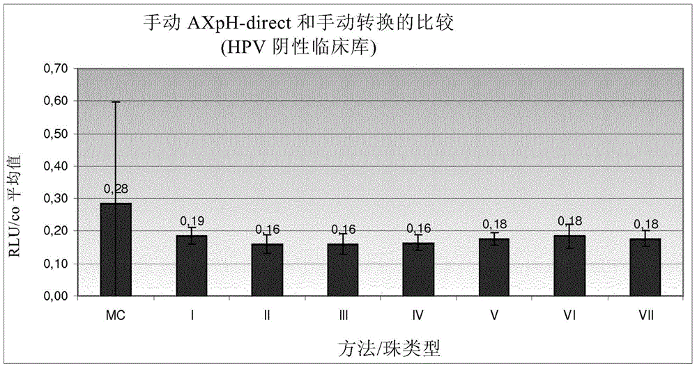
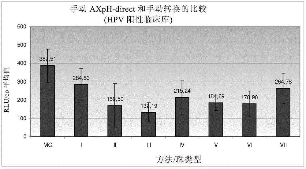
[0380] Example 1: Comparison of manual AXpH-direct and manual conversion

[0381] 1.1) Reference: Sample preparation by manual conversion (MC)

[0382] In MC, which is a standard prior art method, sample preparation is as follows: Samples of HPV-negative clinical pools and HPV-positive clinical pools formulated in culture medium (see below). Obtained by density gradient centrifugation Post-gradient samples, corresponding obtained samples were further processed as described below.

[0383] First, vortex the sample thoroughly. 2.8ml of each sample was transferred to a 15ml centrifuge tube and centrifuged at 800g for 10 minutes. After centrifugation, the supernatant was decanted and the centrifuge tube (opening facing down) was tapped 3 times on a cloth. 200 μl of sample transport medium (Specimen Transport Medium-STM, Digene - containing chaotropic agent) was added to each pellet, ie sample, and vortexed at maximum speed for 15 seconds until all pellets were resuspended...

Embodiment 2

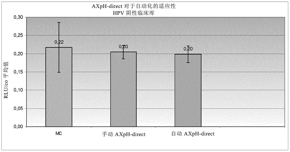
[0400] Example 2: AXpH-direct method for the applicability of automation

[0401] In Example 2, the following samples were processed:

[0402] 1) HPV-negative clinical library in medium (RLU / CO about 0.2),

[0403] 2) SiHa cells in medium (HPV positive; 1 × 10 5 cells / ml),

[0404] 3) HPV-positive clinical pool 1 in medium (RLU / CO approximately 186.4), and

[0405] 4) HPV positive clinical pool 2 in medium (RLU / CO ~530).

[0406] 1.1) Reference: Manual Conversion (MC)

[0407] See Example 1 for the manual conversion (MC) protocol.

[0408] 1.2) Manual AXpH-direct method

[0409] First, vortex the sample thoroughly. Combine 2.8ml of each sample, i.e., with 1.4ml 1.4ml of the original sample mixed with medium, transferred to a 5ml PP-tube. 50 [mu]l of Bead Suspension I (see Example 1) was added. Secure the tube with the cap, carefully invert 10 times, and vortex for 30 seconds. Samples were then incubated at room temperature for 5 minutes. After incubation...


PUM

PropertyMeasurementUnit
sizeaaaaaaaaaa
sizeaaaaaaaaaa
sizeaaaaaaaaaa

Abstract

A method of determining the presence or absence of a target nucleic acid in a cell sample, said method comprising: a) contacting a surface comprising anion exchange moieties with the sample under conditions suitable to induce binding between the cells and said surface; b) separating the surface with the bound cells from the remaining sample to collect the cells; c) releasing nucleic acids from the cells and d) generating a hybrid between the target nucleic acid and a probe specific for the target nucleic acid e) detecting the presence or absence of the hybrid. The present invention is provides a rapid and automatable method, wherein cells, such as epithelial cells originating from cervical swab samples, are collected from the surrounding liquid medium, such as a liquid based cytology medium, by binding them to an anion exchange surface. The cells bind directly with high affinity and quick kinetics to the anion exchange surface which preferably is provided by magnetic particles carrying anion exchange moieties. The cells that are bound to the anion exchange surface can be easily separated from the surrounding medium and can be directly resuspended in a composition that is suitable for a subsequent hybrid capturing assay which is performed in step d) to detect e. g. pathogen nucleic acids such as HPV nucleic acids.

Description

field of invention [0001] The present invention relates to a method for detecting the presence or absence of a target nucleic acid in a liquid sample containing cells. Background of the invention [0002] To detect the presence or absence of a target nucleic acid, several methods are known in the prior art based on capturing hybrids comprising the target nucleic acid to be detected, referred to herein as hybrid capturing assays. The basis of each technique is described, for example, in WO 93 / 10263 and further developments are described in WO 2010 / 127223. Techniques for the detection and characterization of specific nucleic acid sequences and sequence alterations, such as detecting the presence or absence of viral or bacterial nucleic acid sequences indicative of infection, the presence or absence of allelic variants of mammalian genes associated with disease and cancer, and forensic samples Identification of the source of nucleic acids found in , and fetal diagnostics, such...

Claims


Application Information

Patent Timeline
Patent Type & Authority Applications(China)
IPC IPC(8): C12Q1/68C12N15/10
CPCC12N15/101C12N15/1013C12Q1/6806C12Q2563/143C12Q1/6886C12Q1/708C12Q2600/118C12Q2600/158
Inventor M·斯普林格-豪瑟斯C·库普弗P·沙茨
Owner QIAGEN GMBH
Why Eureka
Try Eureka