
Medical image diagnostic device and medical image processing device

A medical image and diagnostic device technology, which is applied in image data processing, medical automatic diagnosis, and equipment for radiological diagnosis, etc. It can solve the problems of low diagnostic ability and low magnification

Active Publication Date: 2015-08-26
TOSHIBA MEDICAL SYST CORP
View PDF9 Cites 5 Cited by
  • Summary
  • Abstract
  • Description
  • Claims
  • Application Information

AI Technical Summary

Problems solved by technology

[0008] However, since the reconstruction of the medical image enlarged by the zoom function is performed at a low magnification ratio, even if the magnification ratio is changed, the roughness of the medical image does not change from that before the image is enlarged.
That is, in the medical image enlarged by the zoom function, the diagnostic ability is low compared with the image subjected to the above-mentioned enlarged reconstruction

Method used


Image

  • Medical image diagnostic device and medical image processing device
  • Medical image diagnostic device and medical image processing device
  • Medical image diagnostic device and medical image processing device

Examples


Embodiment Construction

[0017] Embodiments will be described below with reference to the drawings.

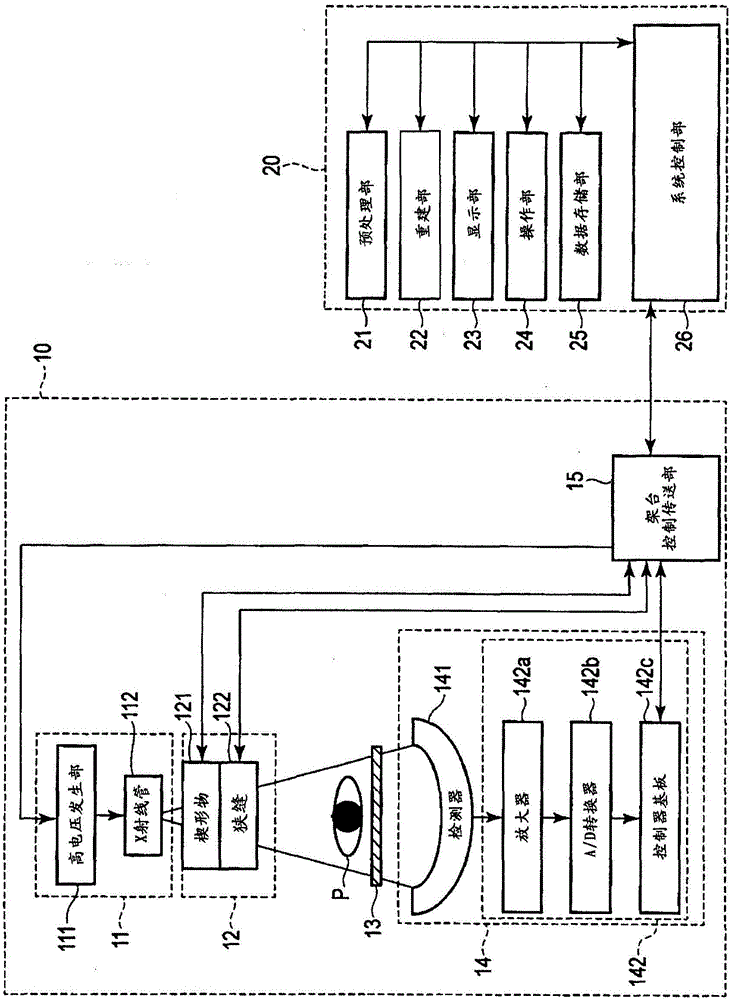
[0018] The medical image diagnostic apparatus according to this embodiment includes, for example, an X-ray computed tomography apparatus (X-ray CT apparatus), a magnetic resonance imaging apparatus (MRI apparatus), an X-ray diagnostic apparatus, and an ultrasonic diagnostic apparatus. In the following description, an X-ray CT apparatus will be described as an example of the medical image diagnosis apparatus according to the present embodiment.

[0019] figure 1 The configuration of the X-ray CT apparatus according to this embodiment is shown. Such as figure 1 As shown, the X-ray CT apparatus includes a gantry 10 and a console 20 .

[0020] The gantry 10 includes an X-ray system 11 , an optical system 12 , a top plate (bed) 13 , a detection system 14 , and a gantry control transmission unit 15 .

[0021] The X-ray system 11 includes a high voltage generator 111 and an X-ray tube 112 . The high vol...


PUM


Abstract

A medical image diagnostic device comprises: an collection unit (10) which collects data relating to the inside of a subject; a reconstruction unit (22) which reconstructs a first image relating to the subject on the basis of the collected data; a display unit (23); a display control unit (26) which displays the reconstructed first image in a display area of the display unit and enlarges and displays a portion of the first image in the display area according to an enlarge display operation by an operator; a range setting unit (26) which sets the displayed enlarged portion of the first image to an enlarged reconstruction range; an enlarge reconstruction unit (22) which enlarges and reconstructs a second image that corresponds to the set enlarged reconstruction range on the basis of a portion of the collected data; and the second image is displayed in place of the portion of the first image in the display area of the display unit by the control of the display control unit.

Description

technical field [0001] Embodiments of the present invention relate to a medical image diagnosis device and a medical image processing device. Background technique [0002] Generally, in the medical field, a subject is diagnosed using medical images obtained by a medical image diagnosis apparatus. Such medical image diagnosis technology is indispensable in current medicine, but it is desired to further improve the diagnostic ability. [0003] Therefore, for example, it is known to perform magnified reconstruction (zoomed reconstruction) in an X-ray computed tomography apparatus (hereinafter referred to as an X-ray CT apparatus) in order to improve diagnostic capabilities. Based on this enlarged reconstruction, a high-resolution image (image with high spatial resolution) is reconstructed by limiting the reconstruction range to a part of the scanning range. By performing such zooming-reconstruction (zooming-reconstruction), a high-resolution image corresponding to the reconst...

Claims


Application Information

Patent Timeline
Patent Type & Authority Applications(China)
IPC IPC(8): A61B6/03
CPCA61B6/469G06T3/40A61B6/032A61B6/5211A61B6/463A61B5/0037A61B5/748G01R33/5608A61B5/055G16H50/20A61B8/14A61B8/463A61B2576/00G06T7/0012G06T2207/10081G06T2207/10088G06T2207/10116
Inventor 山崎正彦佐藤滉一栗原崇加藤祐树渡边孝仁
Owner TOSHIBA MEDICAL SYST CORP
Learn More