

Fully-automatic three-dimensional liver segmentation method based on convolution nerve network
A convolutional neural network, fully automatic technology, used in image analysis, image data processing, instruments, etc., can solve the problems of long segmentation time, low contrast and weak boundaries, over-segmentation, etc., to avoid under-segmentation and over-segmentation, accurate The effect of liver segmentation results
- Summary
- Abstract
- Description
- Claims
- Application Information
AI Technical Summary
Problems solved by technology
Method used
Image

Examples
Embodiment Construction
[0030] Below in conjunction with accompanying drawing and specific embodiment the present invention is described in further detail:
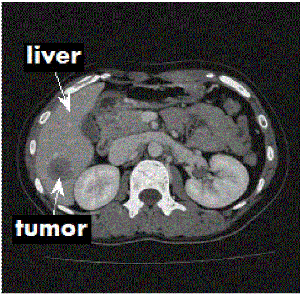
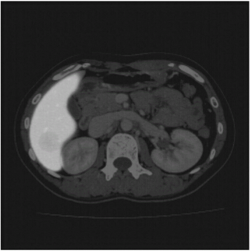
[0031] Provide a new fully automatic 3D liver segmentation method based on convolutional neural network, which is used to segment abdominal liver CTA (Computed Tomography Angiography, CT angiography) volume data, that is, the liver in computerized tomography angiography images, Include the following procedures:
[0032] 1. Prepare the training set;
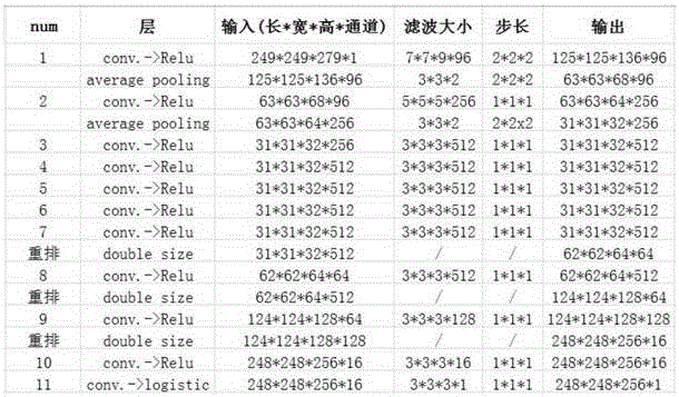
[0033] 2. Training convolutional neural network;
[0034] 3. Use the trained convolutional neural network to process the abdominal liver CTA volume data to obtain the liver segmentation results.
[0035] Described process one specifically comprises the following steps:
[0036] Step A: Collect 68 abdominal liver CTA volume data with a size of 512×512×N, and provide the liver segmentation standard results of these data by doctors and experts, where N is the number of layers of volume data.
[003...
PUM
 Abstract
Description
Claims
Application Information