Determination of time-dependent contrast agent injection curves based on CT scan parameters

A contrast agent and time technology, which is used in hypodermic injection devices, computed tomography scanners, drug devices, etc., can solve problems such as time matching without contrast, non-optimal contrast, etc.

Active Publication Date: 2021-03-09
SIEMENS AG +1
View PDF4 Cites 0 Cited by
  • Summary
  • Abstract
  • Description
  • Claims
  • Application Information

AI Technical Summary

Problems solved by technology

In the case of conventional matching with the aid of a test bolus or bolus tracking, usually only the optimal start time for CT imaging is determined, but there is no temporal matching of the contrast during the imaging process
This therefore produces a non-optimal contrast in at least a sub-region of the region to be imaged of the object under examination

Method used


Image

  • Determination of time-dependent contrast agent injection curves based on CT scan parameters
  • Determination of time-dependent contrast agent injection curves based on CT scan parameters
  • Determination of time-dependent contrast agent injection curves based on CT scan parameters

Examples


Embodiment Construction

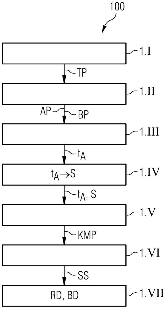
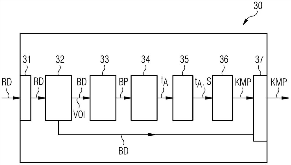
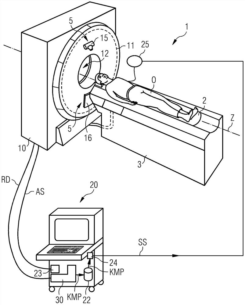
[0031] figure 1 is a flowchart illustrating a method 100 for contrast agent enhanced medical imaging according to an exemplary embodiment of the present invention. The method is described, but not limited to, in conjunction with contrast agent-enhanced imaging by means of a CT system. In step 1.I of the method 100, a topogram TP of the patient's region of interest is first recorded. The internally stored information position map TP can eg be used to determine the position of the patient and the position and size of the various body regions to be examined.

[0032] Then, define the CT inspection protocol in step 1.II. A CT examination protocol defines, for example, a region VOI (also referred to as a scan region) of the patient's body to be imaged. Also defined herein are so-called scan parameters AP, such as for example the rotation time of the rotation system comprising the X-ray source or X-ray tube of the CT system and the detector, the collimation of the X-rays of the C...


PUM


Abstract

A method (100) for automatically determining a contrast agent protocol (KMP) for a contrast agent enhanced medical imaging method is described. The method (100) comprises: overview recording (TP) a region of interest (VOI) of an inspected object (O). In addition, a picture recording protocol (BP) including a plurality of recording parameters (AP) is defined on the basis of the profile recording (TP). Furthermore, on the basis of the image recording protocol (BP), the recording time points (t A ). In addition, the structures acquired using the profile recording (TP) are assigned to the determined recording time points (t A ). Finally, a contrast agent protocol (KMP) including the time course of the contrast agent injection curve (KM) was defined for the acquired structures and the assigned recording time points (t A )The function. A contrast agent protocol determination device (30) for automatically determining a contrast agent protocol (KMP) for a contrast agent-enhanced medical imaging method is also described. Furthermore, an imaging medical device (1) is described.

Description

technical field [0001] The invention relates to a method for determining a time-dependent contrast agent injection profile from CT scan parameters. The invention also relates to a method for performing imaging of a region of interest of an object under examination. The invention also relates to a device for determining a contrast medium injection curve. Finally, the invention relates to a computer tomography system. Background technique [0002] Modern imaging methods are used to generate two-dimensional or three-dimensional image data which can be used to visualize the imaged object to be examined and, additionally, also for further applications. [0003] Imaging methods are often based on the acquisition of x-rays, in which so-called projection measurement data are generated. For example, projection measurement data may be acquired by a computed tomography system (CT system). In the case of using a CT system, usually the combination of an X-ray source and an oppositely...

Claims


Application Information

Patent Timeline
Patent Type & Authority Patents(China)
IPC IPC(8): A61B6/03
CPCA61B6/032A61B6/461A61B6/466A61B6/481A61B6/504A61B6/52A61B6/486A61B6/488A61B6/503A61B6/5217A61B6/541A61B6/469A61M5/007A61M2205/3334A61M2230/04G01R33/5601
Inventor T·弗洛尔G·约斯特H·皮奇
Owner SIEMENS AG
Why Eureka
Try Eureka