In vitro biliary excretion assay

一种胆汁排泄、胆汁的技术,应用在体外胆汁排泄测定法领域,能够解决低精确性等问题

Inactive Publication Date: 2017-09-26
HUREL CORP
View PDF3 Cites 0 Cited by
  • Summary
  • Abstract
  • Description
  • Claims
  • Application Information

AI Technical Summary

Problems solved by technology

The second disadvantage is that bile accumulation of chemical entities is necessarily calculated in double culture assays by measuring two values ​​that are not biliary accumulation, total cellular accumulation (cytoplasmic and biliary) and cytoplasmic accumulation, and then calculating the difference between these values
This assay format has several disadvantages, including low precision

Method used


Image

  • In vitro biliary excretion assay
  • In vitro biliary excretion assay
  • In vitro biliary excretion assay

Examples


Embodiment 1

[0137] Example 1: Hepatocyte-mesenchymal cell co-culture

[0138] Cryopreserved human hepatocytes were removed from liquid nitrogen and thawed. After thawing, cells were resuspended in culture medium and cell number and cell viability were determined using trypan blue exclusion. stromal cells in CO 2 Passage in the incubator until used for experimental plating. On the day of plating, cells were detached from the plate, washed, and resuspended in culture medium. Cell number and viability were determined using trypan blue exclusion.

[0139] Hepatocytes and stromal cells were seeded into collagen-coated 96-well plates at a density of 30,000 hepatocytes per well. Stromal cells were subjected to growth arrest prior to seeding. Cultures were maintained for 7 days before starting any biliary excretion assays. Cells were stained with CDFDA on day 7 to ensure tubule formation before starting the experiment.

Embodiment 2

[0140] Example 2: Excretion of test compounds into bile canaliculi in hepatocyte-stromal cell co-cultures

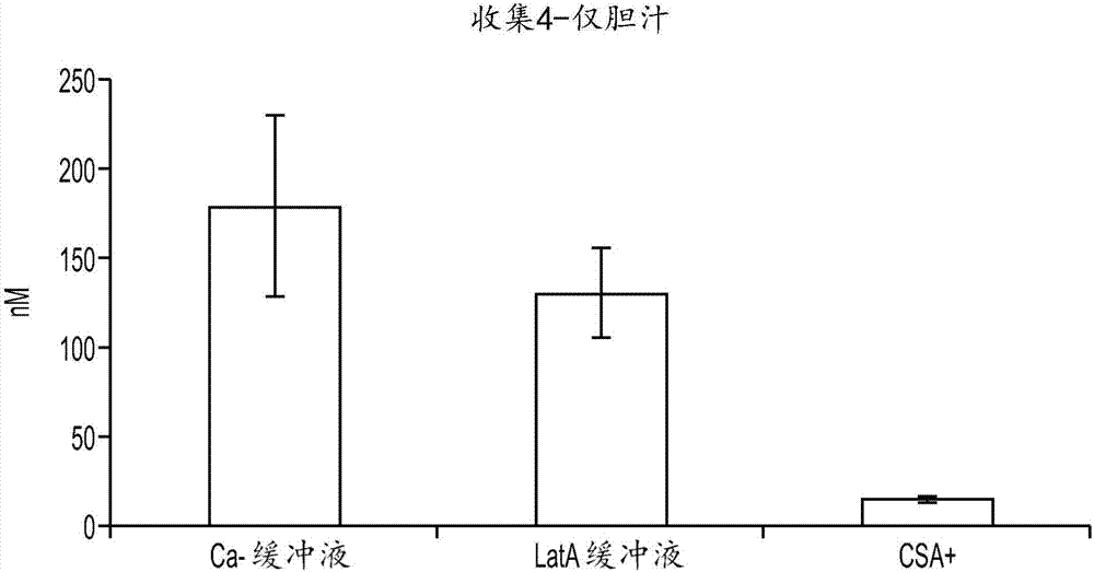
[0141]In this experiment, the presence of bile canaliculi in hepatocyte-stromal cell cultures prepared according to Example 1 was assessed. Co-cultures were incubated with 5 μM 5-(and-6)-carboxy-2',7'-dichloro-fluorescein diacetate (CDFDA) for 20 min. CDFDA uptake by hepatocytes is hydrolyzed by hepatocytes to fluorescent 5-(and-6)-carboxy-2',7'-dichloro-fluorescein (CDA), which is then secreted into tubules in this fluorescent form by the Mrp2 transporter middle. Co-cultures included several tubules that were readily apparent under the microscope (data not shown). Cyclosporin A (CSA) is a known inhibitor of the MrP2 transporter. In subsequent experiments, co-cultures were incubated with CDFDA for 20 min in the presence of 50 μM CSA. After incubation with CDFDA in the presence of CSA, no fluorescent tubules were visible (data not shown). This result demonstrates the...

Embodiment 3

[0142] Example 3: Disruption of tubules

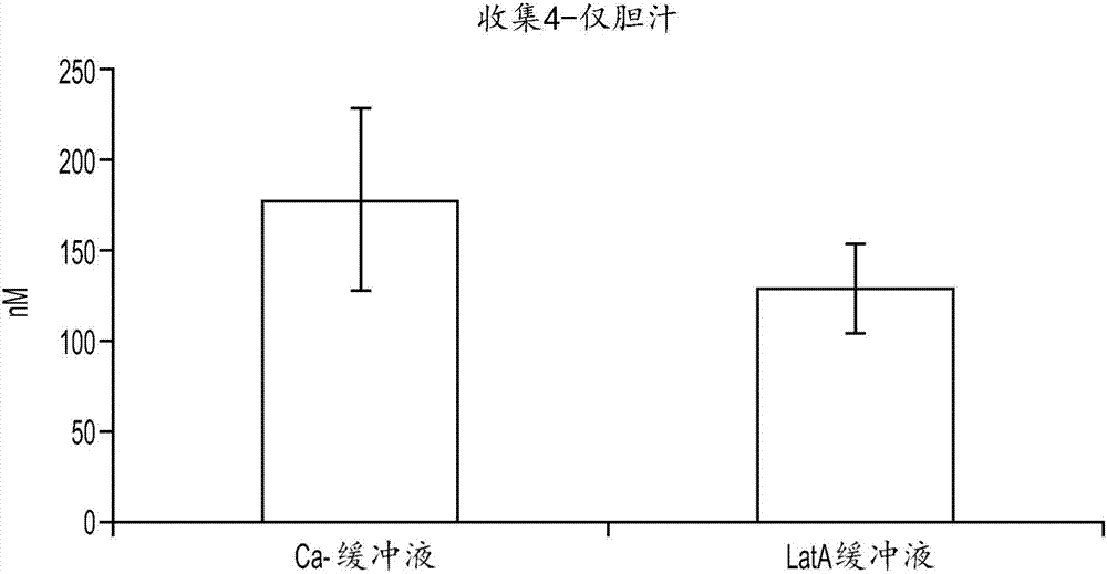
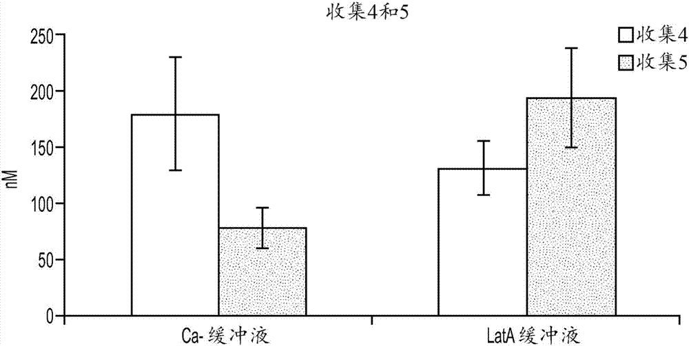
[0143] Assessing the excretion of chemical entities into bile in hepatocyte-stromal cell co-cultures requires a method to distinguish chemical entities present in the tubules of the co-culture from those that may be present in the cytoplasm of cells in the co-culture. Two methods of tubule disruption were tested. In the first method, cultures are exposed to a calcium (Ca-)-free buffer. The lack of calcium inhibits the cells' ability to form tight junctions, which allow bile present in the tubules to escape. First, cells were washed twice in HBSS buffer with Ca+. Cells were then incubated with 5 μM CDFDA in Ca-free HBSS buffer for 20 minutes. Then, cells were washed twice in Ca-free HBSS buffer. No CDF staining was evident as accumulated CDF escaped the tubules after exposure to Ca-buffer (data not shown).

[0144] In the second method, cultures were exposed to a buffer containing Lachunkulin A (LatA). LatA functions to disrupt mi...


PUM


Abstract

An in vitro method of characterizing biliary excretion of a chemical entity using a single hepatocyte culture is disclosed. Comprising providing cell culture comprising hepatocytes forming at least one bile canaliculus; contacting the cell culture with a first chemical entity for a time sufficient to allow uptake of the chemical entity by hepatocytes in the culture; disrupting the at least one bile canaliculus without lysing the hepatocytes and detecting the amount (if any) of the first chemical entity and / or a metabolite thereof released by the at least one bile canaliculus; and lysing the hepatocytes and detecting the amount of the first chemical entity and / or a metabolite thereof released by the hepatocytes.

Description

Background of the invention [0001] It is often undesirable to remove the therapeutic chemical entity from the animal's circulatory system by first-pass metabolism in the liver. If a chemical entity is taken up by hepatocytes and excreted in bile via bile canaliculi, the chemical entity will never reach its therapeutic target. Transporters endogenous to hepatocytes are responsible for moving substrates across the hepatocyte sinusoidal membrane and into the bile canaliculi. Bile canaliculi are structures in liver tissue that receive excreted components from hepatocytes and transport bile to the common bile duct for removal from the animal. Thus, biliary excretion of substrates is a complex process involving translocation across the sinusoidal membrane, movement via the cytoplasm, and transport across the canalicular membrane. [0002] The understanding that hepatobiliary excretion of the parent drug or its metabolites often plays an important role in the overall clearance of t...

Claims


Application Information

Patent Timeline
Patent Type & Authority Applications(China)
IPC IPC(8): C12N5/071C12Q1/04
CPCG01N33/5067G01N33/5091
Inventor E·诺维克C·曹A·帕雷克R·弗里德曼M·L·亚尔穆什E·普鲁德文斯基
Owner HUREL CORP
Why Eureka
Try Eureka