
Medical imaging method and system
A medical imaging system and medical imaging technology, applied in the directions of image generation, image data processing, 2D image generation, etc., can solve the problem of time-consuming imaging methods, and achieve the effect of improving the speed of imaging reconstruction
Patent Information
- Authority / Receiving Office
- CN · China
- Patent Type
- Patents(China)
- Current Assignee / Owner
- Publication Date
- 2021-02-02
Smart Images
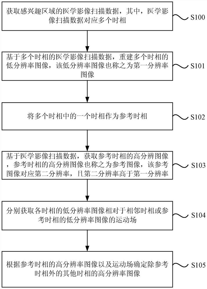
Figure 1 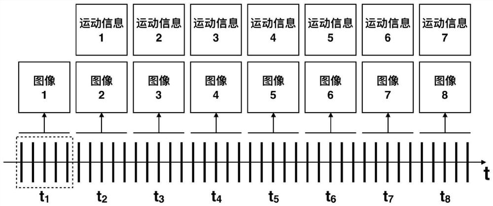
Figure 2 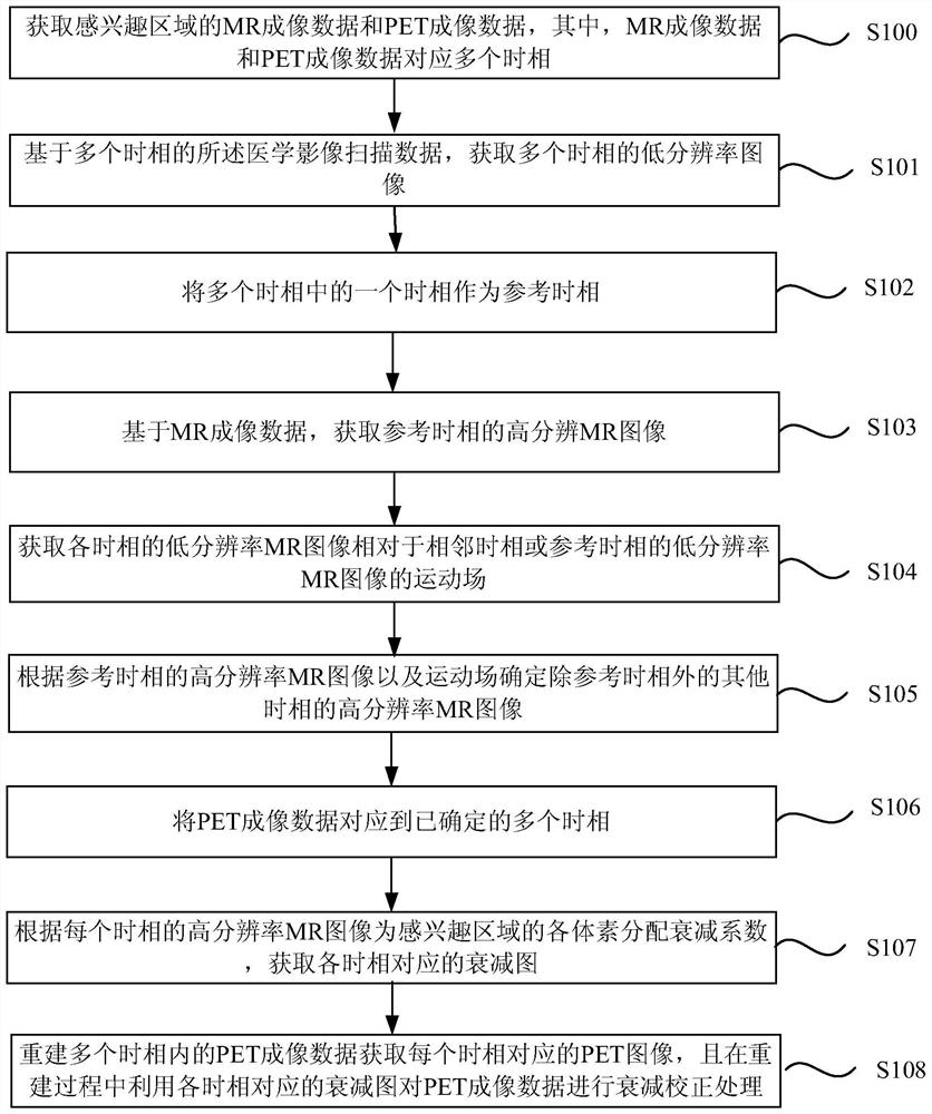
Figure 3
Abstract
Description
technical field
[0001] Embodiments of the present invention relate to image processing technologies, and in particular to a medical imaging method system. Background technique
[0002] Magnetic resonance imaging (Magnetic Resonance Imaging, MRI) utilizes the phenomenon of nuclear magnetic resonance to excite the spin nuclei in human tissues or organs by applying radio frequency pulses, and then receive the electromagnetic signals released by the spin nuclei to reconstruct the image information of human tissue, with ion-free The advantages of radiation, multi-contrast imaging and high soft tissue contrast have become one of the important tools for clinical medical imaging examination. The data collected during the magnetic resonance imaging process is called k-space data, and the magnetic resonance image can be obtained by reconstructing the k-space data. Dynamic imaging obtains a series of time-related k-space data through continuous and repeated scanning of a specific imag...
Examples
Embodiment 1
[0048]figure 1 It is a schematic flow chart of the medical imaging method provided by Embodiment 1 of the present invention, which is applicable to the scene of obtaining dynamic images of target organs or human tissues through medical image scan data of multiple physiological cycles of target organs or human tissues. The medical image scan data is medical image scan data collected by clinical imaging equipment, such as MRI data collected by Magnetic Resonance Imaging (MRI) equipment, CT data collected by Computer Tomography (Computed Tomography, CT) equipment or The PET imaging data collected by positron emission computed tomography (Positron Emission Computed Tomography, PET) equipment may also be imaging data collected by multimodal imaging equipment PET-MR, PET-CT, etc. This embodiment takes MRI data as an example for illustration. The method can be implemented by software or hardware configured in smart devices, such as control computers, personal computers, doctor worksta...
Embodiment 2
[0070] image 3 It is a schematic flow chart of the medical imaging method provided by Embodiment 2 of the present invention. This embodiment is an optimization of the foregoing embodiments, such as image 3 As shown, the method includes:
[0071] S100. Acquire MR imaging data and PET imaging data of the region of interest, where the MR imaging data and PET imaging data correspond to multiple time phases.
[0072] In this embodiment, the medical image scan data may include MR imaging data obtained by scanning the region of interest by the MR scanner during the physiological cycle, and PET imaging data obtained by scanning the region of interest by the PET scanner during the physiological cycle. Optionally, MR imaging data and PET data may be acquired simultaneously, or MR imaging data may be acquired during a PET scan.
[0073] S101. Acquire low-resolution images of multiple time phases based on the medical image scan data of multiple time phases.
[0074] Optionally, at le...
Embodiment 3
[0098] Figure 4 It is a schematic block diagram of a medical imaging device provided in Embodiment 3 of the present invention, which can be implemented by software or hardware configured in smart devices, such as personal computers, doctor workstations, and cloud servers, etc., such as Figure 4 As shown, the imaging device described in this embodiment includes:
[0099] A medical image scan data acquisition module 11, configured to acquire medical image scan data of the region of interest, wherein the medical image scan data corresponds to multiple time phases;
[0100] A low-resolution image acquisition module 12, configured to acquire low-resolution images of multiple time phases based on the medical image scan data of multiple time phases;
[0101] A reference phase determination module 13, configured to use one of the multiple phases as a reference phase;
[0102] A high-resolution image acquisition module 14, configured to acquire a high-resolution image of the referenc...