
Medical image display method, device and computer storage medium

A medical image and display method technology, which is applied in the field of medical images, can solve problems such as the inconvenience of reading images, and achieve the effect of reducing the frequency of switching back and forth

Active Publication Date: 2020-12-04
SHANGHAI UNITED IMAGING HEALTHCARE
View PDF8 Cites 0 Cited by
  • Summary
  • Abstract
  • Description
  • Claims
  • Application Information

AI Technical Summary

Problems solved by technology

However, choosing a different combination of window width and level means that different window width and level values ​​need to be switched back and forth, which brings inconvenience to the reading work

Method used


Image

  • Medical image display method, device and computer storage medium
  • Medical image display method, device and computer storage medium
  • Medical image display method, device and computer storage medium

Examples


Embodiment Construction

[0031] In order to more clearly illustrate the technical solutions of the embodiments of the present application, the following briefly introduces the drawings that need to be used in the description of the embodiments. Obviously, the accompanying drawings in the following description are only some examples or embodiments of the present application, and those skilled in the art can also apply the present application to other similar scenarios. Unless otherwise apparent from context or otherwise indicated, like reference numerals in the figures represent like structures or operations.

[0032] As indicated in this application and claims, the terms "a", "an", "an" and / or "the" do not refer to the singular and may include the plural unless the context clearly indicates an exception. Generally speaking, the terms "comprising" and "comprising" only suggest the inclusion of clearly identified steps and elements, and these steps and elements do not constitute an exclusive list, and ...


PUM


Abstract

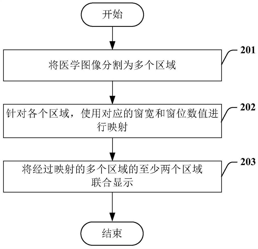
The invention provides a medical image display method and device and a computer storage medium. The medical image display method includes the steps of: dividing a medical image into a plurality of regions; performing mapping for each region using a corresponding window width and window level value; and performing combined display of at least two different regions of the plurality of mapped regions.

Description

technical field [0001] The invention relates to the field of medical images, in particular to a medical image display method, device and computer storage medium. Background technique [0002] Medical images, especially computed tomography (CT, Computed Tomography), positron emission tomography (PET, Positron Emission Tomography) and magnetic resonance images (MR, Magnetic Resonance) are very important medical images, which can non-invasively provide patients with Anatomical structure images, thus providing effective technical support for the diagnosis of related diseases. [0003] The information collected by medical imaging equipment such as CT, PET, and MR is converted into a grayscale image that can be recognized by the naked eye after going through image reconstruction algorithms, image preprocessing and other steps. The original signal intensity value collected by the imaging device has a wide distribution range (such as 0 to 65535), and due to the limitation of the ha...

Claims


Application Information

Patent Timeline
Patent Type & Authority Patents(China)
IPC IPC(8): G06T7/11G16H30/00G16H40/63
CPCG06T2207/10081G06T2207/10088G06T2207/10104G06T7/11
Inventor 荣成城
Owner SHANGHAI UNITED IMAGING HEALTHCARE
Learn More