
Medical image display method, device and storage medium

A medical image and display method technology, applied in the field of image processing, can solve problems such as low processing efficiency, and achieve the effect of speeding up diagnosis efficiency

Active Publication Date: 2021-02-19
SHANGHAI UNITED IMAGING HEALTHCARE
View PDF3 Cites 0 Cited by
  • Summary
  • Abstract
  • Description
  • Claims
  • Application Information

AI Technical Summary

Problems solved by technology

[0004] Based on the problem of low processing efficiency caused by manually adjusting the perspective view in the traditional technology, the purpose of the present invention is to provide a medical image display method, device and storage medium

Method used


Image

  • Medical image display method, device and storage medium
  • Medical image display method, device and storage medium
  • Medical image display method, device and storage medium

Examples


Embodiment Construction

[0029] In order to make the technical solution of the present invention clearer, the technical solution of the present invention will be further described in detail below in conjunction with the accompanying drawings. It should be understood that the specific embodiments described here are only used to explain the present invention and not to limit the present invention. It should be noted that, in the case of no conflict, the embodiments in the present application and the features in the embodiments can be combined with each other.

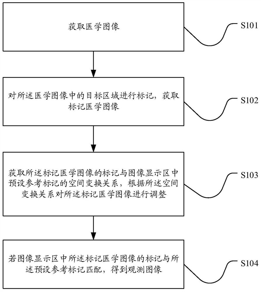
[0030] figure 1 What is shown is a flowchart of a medical image display method provided by an embodiment of the present invention. The method comprises the steps of:

[0031] S101, acquiring a medical image.

[0032] The medical image refers to the image data of the patient taken by a medical instrument. The internal condition of the patient can be clearly displayed on the medical image, which is convenient for medical personnel to analyze the...


PUM


Abstract

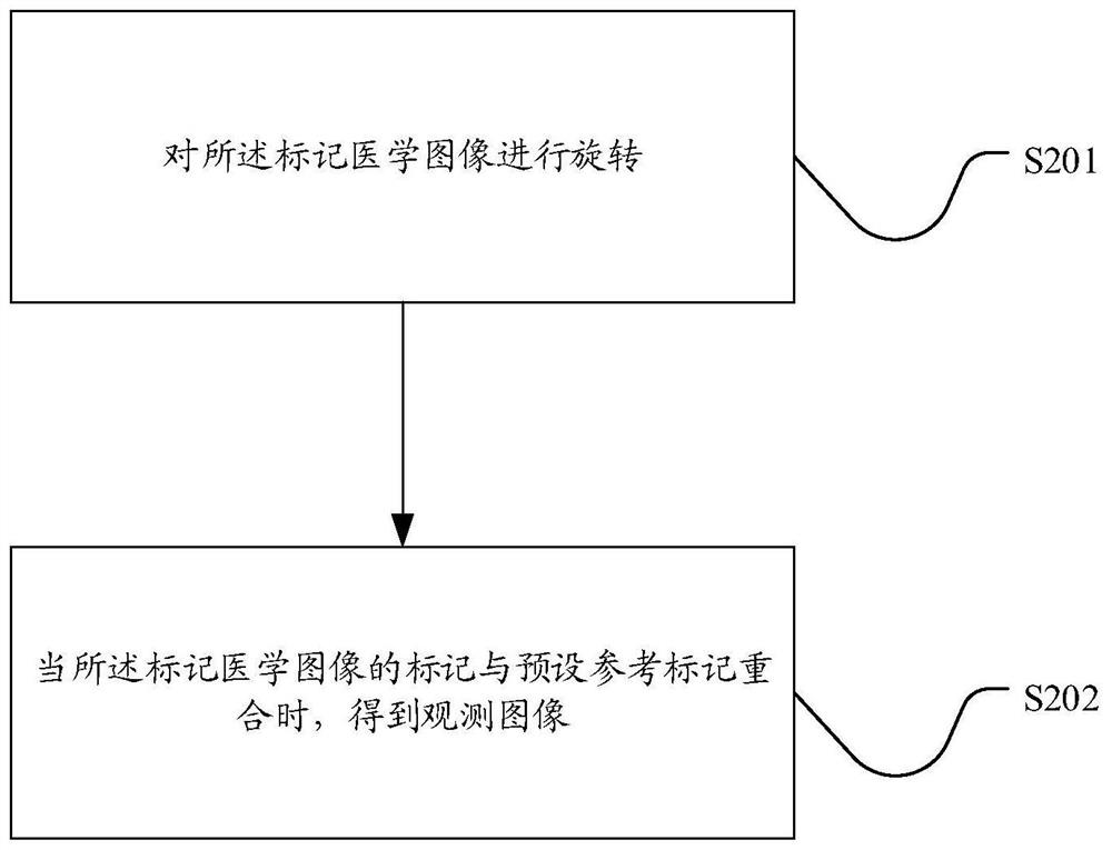
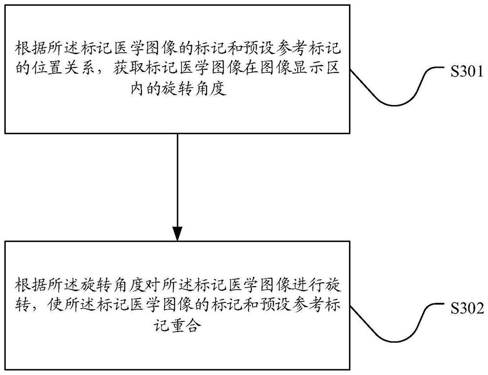
The invention relates to a medical image display method, device and storage medium. Firstly, a medical image is obtained, and target areas in the medical image are marked to obtain the marked medical image, which is convenient for medical personnel to clearly observe prominent structures and parts in the medical image. Acquiring the spatial transformation relationship between the marker of the marked medical image and the preset reference marker in the image display area, and adjusting the marked medical image according to the spatial transformation relationship, if the marker of the marked medical image in the image display area and The preset reference markers are matched to obtain the best observation position of the medical image, so as to speed up the diagnosis efficiency of medical personnel.

Description

technical field [0001] The present invention relates to the field of image processing, in particular to a medical image display method, device and storage medium. Background technique [0002] With the progress of the times, people's demand for medical diagnosis is gradually increasing, such as computerized tomography equipment (CT, Computed Tomography), PET (Positron Emission Tomography) equipment, or a SPECT (Single-Photon-Emission Computed Tomography) equipment, or It is an imaging device such as MRI (Magneticresonance imaging) that is frequently used to obtain medical images. After performing medical imaging, doctors need to adjust the medical image to a specified angle to facilitate observation of the medical image. [0003] In the traditional technology, the doctor adjusts the medical image to the specified observation orientation through manual rotation and translation operations, and it is a continuous and progressive manual operation, gradually approaching the spec...

Claims


Application Information

Patent Timeline
Patent Type & Authority Patents(China)
IPC IPC(8): G06T7/73G06T3/60
CPCG06T3/608G06T2207/30004G06T7/74
Inventor 聂卫文于文君
Owner SHANGHAI UNITED IMAGING HEALTHCARE
Learn More