Sarcoidosis benign and malignant prediction method based on ResNet-Inception model

A prediction method and technology of pulmonary nodules, applied in the field of medical image processing, can solve problems such as insufficient robustness and local areas

Active Publication Date: 2018-05-11
NORTHEASTERN UNIV
View PDF3 Cites 19 Cited by
  • Summary
  • Abstract
  • Description
  • Claims
  • Application Information

AI Technical Summary

Problems solved by technology

However, the main disadvantage of the traditional algorithm is that the extracted features are only local areas, which are not robust enough for common variance and other features.

Method used


Image

  • Sarcoidosis benign and malignant prediction method based on ResNet-Inception model
  • Sarcoidosis benign and malignant prediction method based on ResNet-Inception model
  • Sarcoidosis benign and malignant prediction method based on ResNet-Inception model

Examples


Embodiment Construction

[0028] The specific embodiments of the present invention will be described in detail below in conjunction with the accompanying drawings.

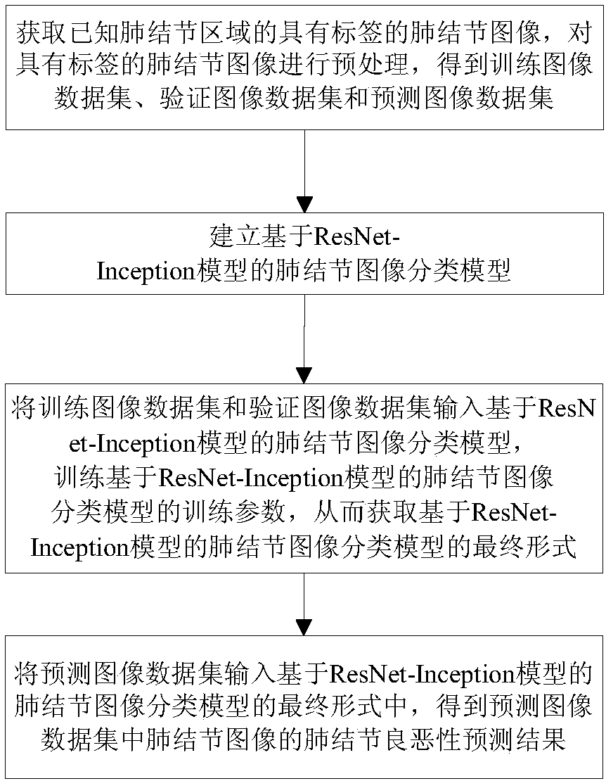
[0029] A method for predicting benign and malignant pulmonary nodules based on the ResNet-Inception model, such as figure 1 shown, including the following steps:

[0030] Step 1: Acquire labeled pulmonary nodule images of known pulmonary nodule regions, preprocess the labeled pulmonary nodule images, and obtain training image datasets, verification image datasets, and predicted image datasets.

[0031] Step 1.1: Obtain a labeled pulmonary nodule image of a known pulmonary nodule region, perform nodule region segmentation on the labeled pulmonary nodule image, and perform cropping to obtain a cropped pulmonary nodule image.

[0032] In this embodiment, 700 labeled pulmonary nodule images of known pulmonary nodule regions are obtained from the Lung Image Database Consortium (LIDC-IDRI). According to the real labels of four radiologists, th...


PUM


Abstract

The present invention provides a sarcoidosis benign and malignant prediction method based on a ResNet (Residual Network)-Inception model. The method comprises the steps of: obtaining a sarcoidosis image with a tag of a known sarcoidosis area, performing preprocessing of the sarcoidosis image with the tag, and obtaining a training image data set, a verification image data set and a prediction imagedata set; establishing a sarcoidosis image classification model based on a ResNet-Inception model; and inputting the predication image data set into a final mode of the sarcoidosis image classification model based on the ResNet-Inception model, and obtaining a sarcoidosis benign and malignant prediction result of the sarcoidosis image in the prediction image dataset. The method designs a new network structure model, and can obtain sarcoidosis benign and malignant prediction according to a sarcoidosis CT image.

Description

technical field [0001] The invention belongs to the technical field of medical image processing, and in particular relates to a method for predicting benign and malignant pulmonary nodules based on a ResNet-Inception model. Background technique [0002] The lesions of lung diseases on medical imaging usually appear as pulmonary nodules. According to the continuous research of pattern recognition and other methods, people have proposed the technology of using computers to assist radiologists to detect pulmonary nodules, that is, Computer Aided Diagnosis (Computer Aided Diagnosis). Diagnosis, CAD) system. In CAD, it involves evaluation or classification with some common algorithms, such as algorithms such as neural networks, k-means or support vector machines. However, the main disadvantage of the traditional algorithm is that the extracted features are only local areas, which are not robust enough for common variance and other features. Based on the differences of data sets...

Claims


Application Information

Patent Timeline
Patent Type & Authority Applications(China)
IPC IPC(8): G16H50/20G06T7/11G06N3/04G06N3/08
CPCG06N3/08G06T7/11G06T2207/30064G06T2207/20132G06N3/045
Inventor 齐守良刘力瑶杨帆赵歆卓张白桦钱唯
Owner NORTHEASTERN UNIV
Why Eureka
Try Eureka