
Automatic segmentation method for lesion area in digital pathological full slice image

A digital pathology and lesion area technology, applied in image analysis, image enhancement, image data processing, etc., can solve the problem of large size of full-slice images, inability to integrate analysis results of multiple image blocks, lack of analysis results of digital pathology full-slice images, etc. problem, to achieve accurate prediction and analysis

Pending Publication Date: 2018-10-09
MOTIC XIAMEN MEDICAL DIAGNOSTICS SYST
View PDF9 Cites 14 Cited by
  • Summary
  • Abstract
  • Description
  • Claims
  • Application Information

AI Technical Summary

Problems solved by technology

[0003] Because the key information such as cell structure and distribution required for the diagnosis of digital pathology full slice images is stored under high-power microscopes, and under this condition, the size of digital pathology full slice images is huge, up to 100,000×100,000 pixels, and full slices cannot be realized with current computer capabilities. overall treatment
In the current application of pathological image analysis, the digital pathological full slice image is generally cut into image blocks for processing, so that the analysis results of multiple image blocks cannot be integrated into the digital pathological full slice image, which leads to the lack of analysis results of the digital pathological full slice image. Cause great difficulty in the realization of assisted diagnosis or automatic diagnosis

Method used


Image

  • Automatic segmentation method for lesion area in digital pathological full slice image
  • Automatic segmentation method for lesion area in digital pathological full slice image
  • Automatic segmentation method for lesion area in digital pathological full slice image

Examples


Embodiment Construction

[0026] The following will clearly and completely describe the technical solutions in the embodiments of the present invention with reference to the accompanying drawings in the embodiments of the present invention. Obviously, the described embodiments are only some, not all, embodiments of the present invention. Based on the embodiments of the present invention, all other embodiments obtained by persons of ordinary skill in the art without making creative efforts belong to the protection scope of the present invention.

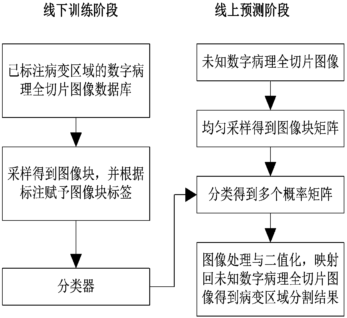
[0027] The embodiment of the present invention discloses a method for automatically segmenting the lesion area of ​​a digital pathological full slice image, such as figure 1 As shown, it is applied to the full-slice database of the marked lesion area and the unknown digital pathological full-slice image, including the offline training stage and the online prediction stage.

[0028] (1) The operation steps in the offline training phase include:

[0029] S11: d...


PUM


Abstract

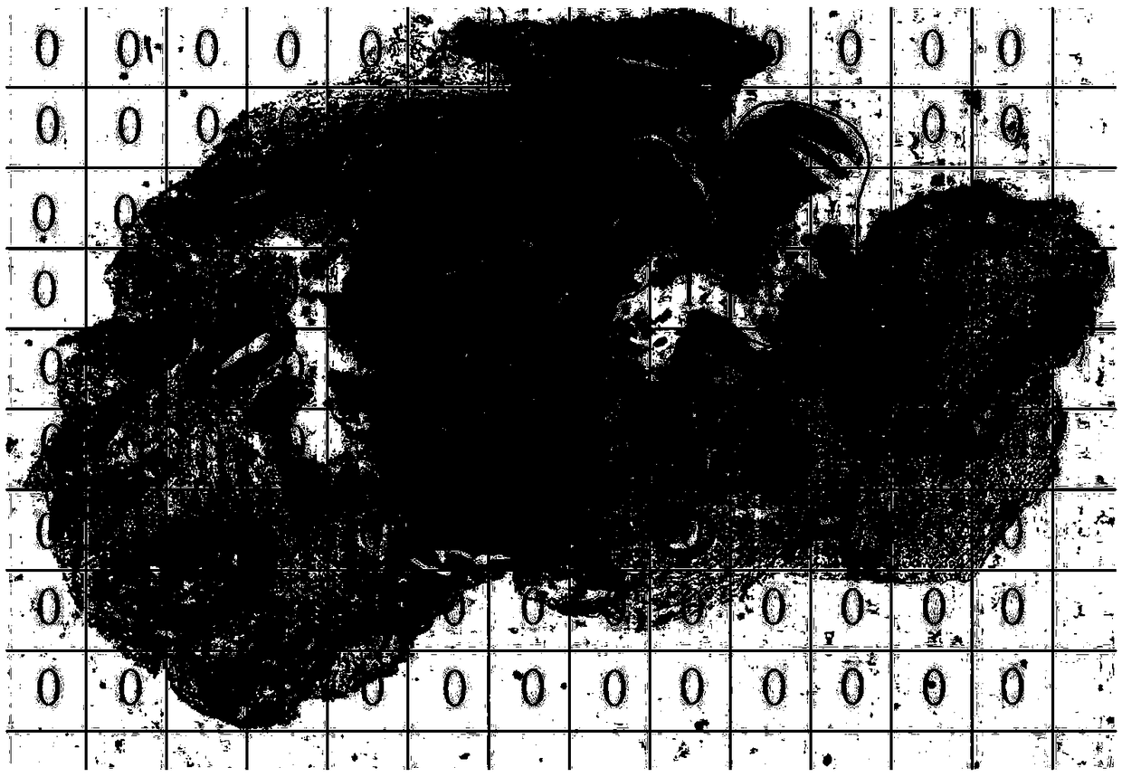
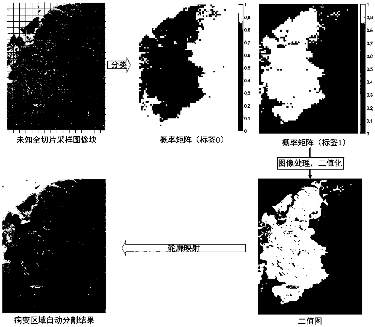
The invention discloses an automatic segmentation method for a lesion area in a digital pathological full slice image. The method comprises steps that an offline training phase and an online prediction phase are included, for the training phase, a digital pathological full slice image in a full slice database with the labeled lesion area is sampled to obtain a large number of labeled image blocks,and a classifier is trained; for the prediction stage, an image block matrix is obtained through uniformly sampling an unknown digital pathological full slice image, multiple probability matrixes areobtained by the classifier, the probability matrixes are processed and binarized, and the obtained contour is mapped back to the unknown digital pathological full slice image to obtain the segmentation result. The method is advantaged in that automatic segmentation of the lesion area of the unknown full slice can be achieved simply through the full-slice database with the labeled lesion area, notonly lesion categories of all the possible lesion areas in the full slice are shown, but also the contour and position distribution of each lesion area are further accurately displayed, and comprehensive, intuitive and accurate prediction and analysis of the unknown full-slice lesion status are realized.

Description

technical field [0001] The present invention relates to the technical field of digital image processing and machine learning, and more specifically relates to a method for automatically segmenting lesion regions of digital pathological whole slice images. Background technique [0002] In recent years, with the rapid development of pathology and computer technology, the number of digital pathology whole slide images has grown rapidly. Digital pathological whole slide images are large-size, high-resolution digital images obtained by scanning and collecting traditional glass pathological slides through automatic microscopes or optical magnification systems, and are an important basis for pathologists in diagnosis. [0003] Because the key information such as cell structure and distribution required for the diagnosis of digital pathology full slice images is stored under high-power microscopes, and under this condition, the size of digital pathology full slice images is huge, up...

Claims


Application Information

Patent Timeline
IPC IPC(8): G06T7/10G06N3/04G06K9/62G06N3/00G06T3/40
CPCG06N3/006G06T3/4038G06T7/10G06T2207/30004G06T2207/20081G06N3/045G06F18/2411
Inventor 姜志国麻义兵郑钰山
Owner MOTIC XIAMEN MEDICAL DIAGNOSTICS SYST
Learn More