An automatic segmentation method for abdominal CT images of liver lesions based on a three-level cascaded network

A CT image and cascade network technology, applied in the field of image processing, can solve the problems of low soft tissue contrast, low contrast, and operator dependence, and achieve the effect of reducing false positives and accurate automatic segmentation

Active Publication Date: 2021-08-13
NORTHEASTERN UNIV LIAONING
View PDF8 Cites 0 Cited by
  • Summary
  • Abstract
  • Description
  • Claims
  • Application Information

AI Technical Summary

Problems solved by technology

In clinical routine, although manual and semi-manual techniques are used, however, these methods are subjective, heavily operator-dependent and very time-consuming
Computer-aided methods have been developed in the past to increase the productivity of radiologists, however due to the low contrast of the liver with its lesions, different types of contrast, abnormalities in the tissue (metastatic resection), size and number of lesions vary , automatic liver and its lesion segmentation is still a very challenging problem
Additionally, CT images typically have low soft tissue contrast and are susceptible to noise and other artifacts
[0003] Existing methods for segmentation of the liver and its lesions based on intensity clustering, region growing, or deformable models have shown limited success in addressing this difficult problem
This complexity of contrasting differences makes it difficult to generalize intensity-based methods to unseen test cases at different clinical points
Furthermore, the varying shape of lesions due to irregular tumor growth and response to treatment (i.e., surgical resection) reduces the efficiency of computational methods that exploit prior knowledge of lesion shape
Therefore several interactive and automatic methods (including gray-scale and texture-based methods, graph-cut methods, level sets, sigmoid edge modeling) that have been developed for segmenting the liver and its lesions in CT volumes are not widely used clinically. application

Method used


Image

  • An automatic segmentation method for abdominal CT images of liver lesions based on a three-level cascaded network
  • An automatic segmentation method for abdominal CT images of liver lesions based on a three-level cascaded network
  • An automatic segmentation method for abdominal CT images of liver lesions based on a three-level cascaded network

Examples


Embodiment Construction

[0072] In order to better explain the present invention and facilitate understanding, the present invention will be described in detail below through specific embodiments in conjunction with the accompanying drawings. The specific implementation manners of the present invention will be described in detail below in conjunction with the accompanying drawings.

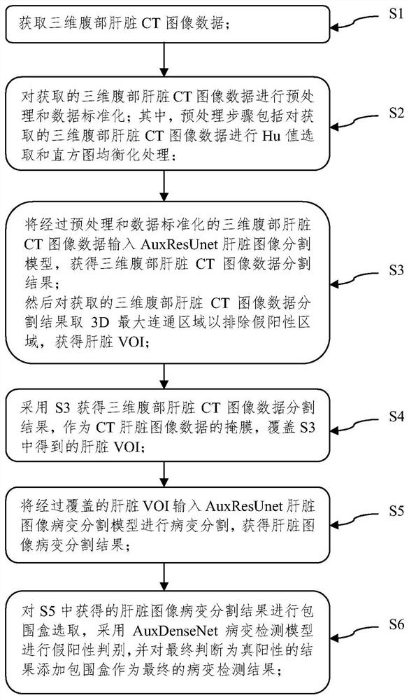
[0073] like figure 1 As shown: this embodiment discloses a method for automatic segmentation of abdominal CT liver lesion images based on a three-level cascaded network, the method comprising:

[0074] S1. Obtain three-dimensional abdominal liver CT image data.

[0075] It should be noted that the three-dimensional abdominal liver CT image data obtained here includes a test set and a training set, where the test set is used to test the performance of the three-level cascaded network, and the training set is used for the three-level cascaded network in this embodiment. Model training.

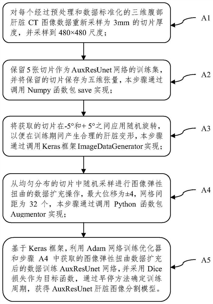
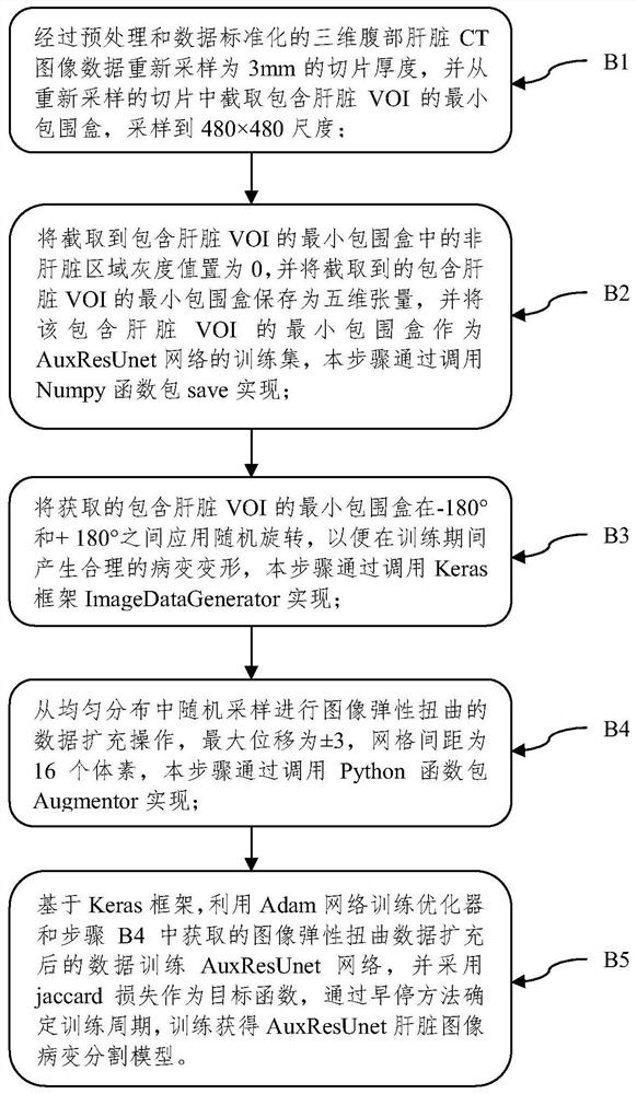
[0076] S2. Perform preprocessing ...


PUM


Abstract

The present invention relates to a method for automatically segmenting abdominal CT liver lesion images based on a three-level cascade network, the method comprising: S1, acquiring three-dimensional abdominal liver CT image data; S2, pre-processing the acquired three-dimensional abdominal liver CT image data Processing and data standardization; S3. Input the preprocessed and data-standardized three-dimensional abdominal liver CT image data into the AuxResUnet liver image segmentation model, and then take the 3D maximum connected area for the obtained three-dimensional abdominal liver CT image data segmentation result to exclude false positive areas , obtain liver VOI; S4, use S3 to obtain the segmentation result of three-dimensional abdominal liver CT image data, use it as a mask of CT liver image data, and cover the liver VOI obtained in S3; S5, input the covered liver VOI into AuxResUnet liver image lesion segmentation The model performs lesion segmentation to obtain liver image lesion segmentation results; the image segmentation method provided by the present invention can realize rapid and accurate segmentation of liver and liver lesions.

Description

technical field [0001] The invention belongs to the technical field of image processing, and in particular relates to an automatic segmentation method for abdominal CT liver lesion images based on a three-level cascade network. Background technique [0002] Abnormal morphology and texture of the liver and visible lesions on CT are important biomarkers of disease progression in both primary and secondary liver neoplastic diseases. In clinical routine, although manual and semi-manual techniques are used, however, these methods are subjective, heavily operator-dependent, and very time-consuming. Computer-aided methods have been developed in the past to increase the productivity of radiologists, however due to the low contrast of the liver with its lesions, different types of contrast, abnormalities in the tissue (metastatic resection), size and number of lesions vary , automatic liver and its lesion segmentation remains a very challenging problem. Additionally, CT images typi...

Claims


Application Information

Patent Timeline
Patent Type & Authority Patents(China)
IPC IPC(8): G06T7/00G06T7/10G06K9/62
CPCG06T7/0012G06T7/10G06T2207/30056G06F18/24
Inventor 姜慧研史天予白志奇黄亮亮
Owner NORTHEASTERN UNIV LIAONING
Why Eureka
Try Eureka