Lung parenchyma segmentation method for extracting CT image based on clustering key frames

A technology of CT images and lung parenchyma, which is applied in the field of medical image processing, can solve the problems of unsatisfactory processing speed and efficiency, long development cycle, and high complexity, and achieve fast and accurate segmentation, reduce the work intensity of doctors, and ensure segmentation accuracy Effect

Pending Publication Date: 2020-05-12
TIANJIN TUMOR HOSPITAL
View PDF7 Cites 4 Cited by
  • Summary
  • Abstract
  • Description
  • Claims
  • Application Information

AI Technical Summary

Problems solved by technology

However, due to the large amount of lung CT data and large individual differences, the existing CAD algorithms have disadvantages such as long development cycle, high complexity, and unsatisfactory processing speed and efficiency.

Method used


Image

  • Lung parenchyma segmentation method for extracting CT image based on clustering key frames
  • Lung parenchyma segmentation method for extracting CT image based on clustering key frames
  • Lung parenchyma segmentation method for extracting CT image based on clustering key frames

Examples


Embodiment Construction

[0026] The present invention will be further described in detail below in conjunction with the embodiments, so that those skilled in the art can implement it with reference to the description.

[0027] It should be understood that terms such as "having", "comprising" and "including" used herein do not exclude the presence or addition of one or more other elements or combinations thereof.

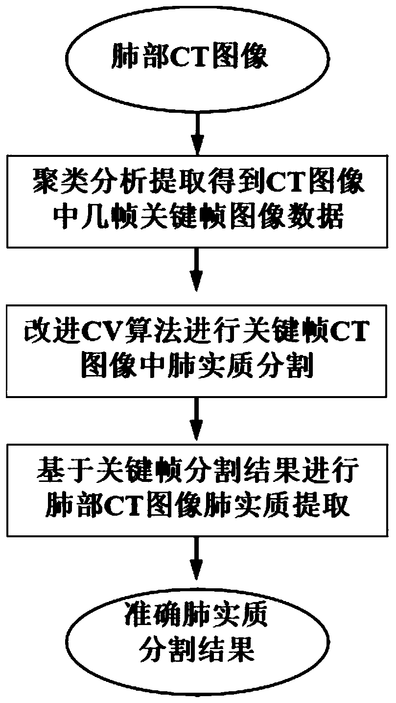
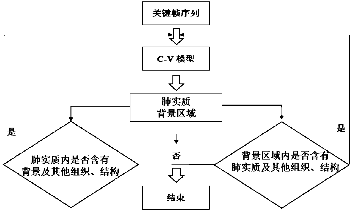
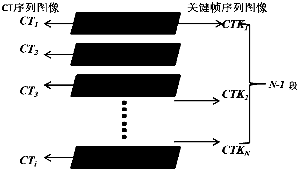
[0028] like figure 1 As shown, a method for fast and automatic lung parenchyma segmentation of CT images based on clustering key frame extraction in this embodiment includes the following steps:

[0029] Step 1, read the CT image of the patient's lungs.

[0030] This step preprocesses the CT image of the patient's lungs, mainly including division of chest cavity regions and image grayscale transformation processing. details as follows:

[0031] (1) Chest area of ​​interest division: Remove the image data other than the bed board, head and neck and other thoracic cavity in the patient's CT...


PUM


Abstract

The invention relates to a lung parenchyma segmentation method for extracting a CT image based on a clustering key frame, and the method comprises the following steps: carrying out the preprocessing of lung CT image data, and carrying out the lung window windowing gray scale transformation of an image CT value; performing clustering analysis on the grey level histogram similarity of the patient lung CT image processed in the step 1, and extracting key frame data in the lung CT sequence; carrying out pulmonary parenchyma segmentation on the patient key frame CT image by adopting a multi-phase level set CV model; performing lung parenchyma mapping segmentation extraction in the lung CT complete sequence according to a lung parenchyma segmentation result in the key frame to obtain a lung parenchyma initial contour; and performing morphological corrosion and expansion operation on the lung parenchyma initial contour in the lung CT image to refine the contour, and finally obtaining a lung parenchyma segmentation result in the patient lung CT data. The invention also provides lung parenchyma segmentation equipment for realizing the segmentation method.

Description

technical field [0001] The invention relates to the field of medical image processing, in particular to a fast and automatic lung parenchymal segmentation method for extracting CT images based on clustering key frames. Background technique [0002] Lung computed tomography (CT) imaging has good tissue density resolution, which can effectively and directly realize non-invasive and low-cost screening of early lung cancer, and has become a clinically recommended detection method. With the continuous improvement of clinical demand for lung imaging accuracy, the thickness of CT scan layers is gradually reduced, resulting in an explosive increase in the amount of clinical lung CT data, which brings problems such as large clinical processing workload, slow speed and low efficiency. . Therefore, the Computer-Aided Diagnosis (CAD) technology developed based on various machine learning algorithms can realize fast and accurate automatic processing and analysis of CT images, which will...

Claims


Application Information

Patent Timeline
Patent Type & Authority Applications(China)
IPC IPC(8): G06T7/11G06T7/136G06T7/194G06K9/62
CPCG06T7/11G06T7/136G06T7/194G06T2207/10081G06T2207/30061G06F18/2321
Inventor 于旭耀王平袁智勇王煜雯余辉宋勇春董洋孙敬来
Owner TIANJIN TUMOR HOSPITAL
Why Eureka
Try Eureka