
Medical image processing method, device, equipment and storage medium

A medical image and processing method technology, applied in the field of medical image processing, can solve the problems of high time cost and poor user experience of three-dimensional images of blood vessels

Active Publication Date: 2021-01-26
QINGDAO HISENSE MEDICAL EQUIP
View PDF10 Cites 0 Cited by
  • Summary
  • Abstract
  • Description
  • Claims
  • Application Information

AI Technical Summary

Problems solved by technology

This makes the time cost of creating 3D images of blood vessels high, and its user experience is poor

Method used


Image

  • Medical image processing method, device, equipment and storage medium
  • Medical image processing method, device, equipment and storage medium
  • Medical image processing method, device, equipment and storage medium

Examples


Embodiment Construction

[0035]In order to make the objectives, technical solutions and advantages of the present invention clearer, the embodiments of the present invention will be described in further detail below in conjunction with the accompanying drawings.

[0036]Before explaining the embodiments of the present invention in detail, the application scenarios of the embodiments of the present invention will be introduced. The method provided in the embodiment of the present invention is applied to a terminal, which is a medical device in a medical scene, or is called a medical device, a medical image processing device, etc., and the medical device may be a display device for medical images. For example, the terminal is a computer-assisted medical display device, such as a computer, a Computed Tomography (CT) machine, an MRI machine, etc., and the medical image may be a three-dimensional medical reconstruction model, etc., which is the embodiment of the present invention. Not limited.

[0037]By executing the...


PUM


Abstract

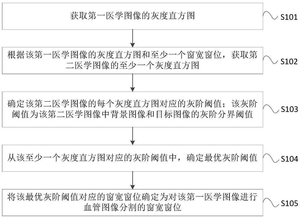
The invention provides a medical image processing method, device, equipment and storage medium. The medical image processing method provided by the present invention includes: acquiring the grayscale histogram of the first medical image; acquiring at least one grayscale of the second medical image according to the grayscale histogram of the first medical image and at least one window width and window level grayscale histogram; determine the grayscale threshold corresponding to each grayscale histogram of the second medical image; determine the optimal grayscale threshold from the grayscale thresholds corresponding to at least one grayscale histogram; The window width and level corresponding to the optimal gray scale threshold is determined as the window width and level for performing blood vessel image segmentation on the first medical image. The invention can shorten the process of determining the window width and level, reduce the time cost of creating a three-dimensional image of blood vessels, and improve user experience.

Description

Technical field[0001]The present invention relates to medical image processing technology, in particular to a medical image processing method, device, equipment and storage medium.Background technique[0002]With the continuous development of medical imaging technology, the use of mathematical models is an important means of assisting various vascular diseases. The rich three-dimensional images of blood vessels play an important role in guiding surgery such as tumor removal.[0003]In the process of creating a three-dimensional image of blood vessels, when a window width and window level is selected, the window width and window level can be used to adjust and display each image in a group of images, so as to display the blood vessel pixels in each image. Distinguish from the background image. However, in certain window widths, the pixel points of the blood vessel may be difficult to distinguish from the background image, which makes the recognition of the blood vessel pixels inaccurate,...

Claims


Application Information

Patent Timeline
Patent Type & Authority Patents(China)
IPC IPC(8): G06T7/00G06T7/136
CPCG06T7/0012G06T2207/30101G06T7/136
Inventor 庞晓磊田广野陈永健
Owner QINGDAO HISENSE MEDICAL EQUIP
Learn More