
Pathological picture identifying method and device

An identification method and picture technology, applied in the medical field, can solve the problems of time-consuming and labor-intensive, different identification conclusions, and unsatisfactory accuracy, and achieve the effect of improving accuracy and efficiency.

Active Publication Date: 2019-02-01
BEIJING CANCER HOSPITAL PEKING UNIV CANCER HOSPITAL +1
View PDF7 Cites 19 Cited by
  • Summary
  • Abstract
  • Description
  • Claims
  • Application Information

AI Technical Summary

Problems solved by technology

The evaluation of lymph node metastasis in pathological diagnosis is the key to diagnosis. Pathologists need to carefully and carefully observe each lymph node one by one. The whole process is not only time-consuming and labor-intensive, but also depends on experience. of doctors may have different identification conclusions for the same pathological picture

Method used


Image

  • Pathological picture identifying method and device
  • Pathological picture identifying method and device
  • Pathological picture identifying method and device

Examples


Embodiment Construction

[0020] In order to make the purpose, technical solutions and advantages of the embodiments of the present invention more clear, the embodiments of the present invention will be further described in detail below in conjunction with the accompanying drawings. Here, the exemplary embodiments and descriptions of the present invention are used to explain the present invention, but not to limit the present invention.

[0021] Before introducing the embodiments of the present invention, the technical terms involved in the present invention are firstly introduced.

[0022] 1. False positive rate: false positive rate, the ratio of the number of samples that are actually negative and predicted positive by the model to all negative samples.

[0023] 2. False negative rate: false negative rate, the ratio of the number of samples that are actually positive and the model predicts negative to all positive samples.

[0024] 3. Accuracy rate: Accuracy=(number of samples predicted correctly) / (...


PUM


Abstract

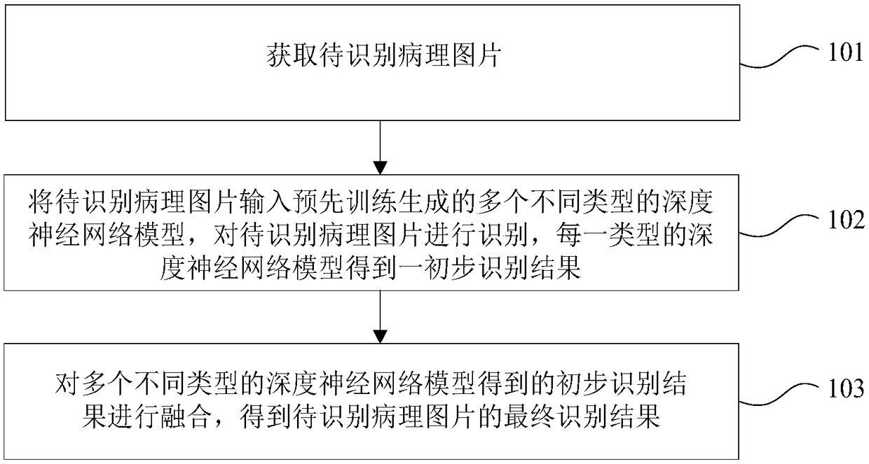
The invention discloses a pathological picture identifying method and device. The method comprises steps: a to-be-recognized pathological picture is acquired; the to-be-recognized pathological pictureis inputted to multiple different types of depth neural network models generated by pre-training, the to-be-recognized pathological picture is identified, and an initial identifying result is obtained by each type of depth neural network model, wherein the multiple different types of depth neural network models are generated through pre-training according to multiple pathological picture samples;and the initial identifying results obtained by the multiple different types of depth neural network models are fused to obtain a final identifying result of the to-be-recognized pathological picture. The above technical scheme improves the pathological picture identifying efficiency and the accuracy.

Description

technical field [0001] The invention relates to the field of medical technology, in particular to a method and device for identifying pathological pictures. Background technique [0002] Lymph node metastasis is the most common way of tumor metastasis. Radical resection for advanced gastric cancer includes complete resection of the primary gastric cancer lesion, metastatic lymph nodes, and invaded tissues and organs. Postoperative pathological diagnosis of gastric cancer is the gold standard for the diagnosis of gastric cancer, which provides an important basis for the staging and treatment of patients. The evaluation of lymph node metastasis in pathological diagnosis is the key to diagnosis. Pathologists need to carefully and carefully observe each lymph node one by one. The whole process is not only time-consuming and labor-intensive, but also depends on experience. Doctors may have different identification conclusions for the same pathological picture. Contents of the...

Claims


Application Information

Patent Timeline
Patent Type & Authority Applications(China)
IPC IPC(8): G16H30/20G16H50/20
CPCG16H30/20G16H50/20
Inventor 孙宇季加孚王鑫宇李元骏李子禹吴晓江步召德
Owner BEIJING CANCER HOSPITAL PEKING UNIV CANCER HOSPITAL
Learn More