A method and system for intercepting intracranial blood vessel images based on a centerline

A technique of intracranial blood vessels and centerline, applied in the field of medical imaging

Active Publication Date: 2019-03-08
UNION STRONG (BEIJING) TECH CO LTD
View PDF27 Cites 8 Cited by
  • Summary
  • Abstract
  • Description
  • Claims
  • Application Information

AI Technical Summary

Problems solved by technology

[0007] However, in the prior art, there is no local interception method

Method used


Image

  • A method and system for intercepting intracranial blood vessel images based on a centerline
  • A method and system for intercepting intracranial blood vessel images based on a centerline
  • A method and system for intercepting intracranial blood vessel images based on a centerline

Examples


Embodiment Construction

[0038] The embodiments of the present application provide a method and system for capturing images of intracranial blood vessels based on a centerline, so as to solve the problem of local capturing of images of blood vessel segments in intracranial blood vessel images.

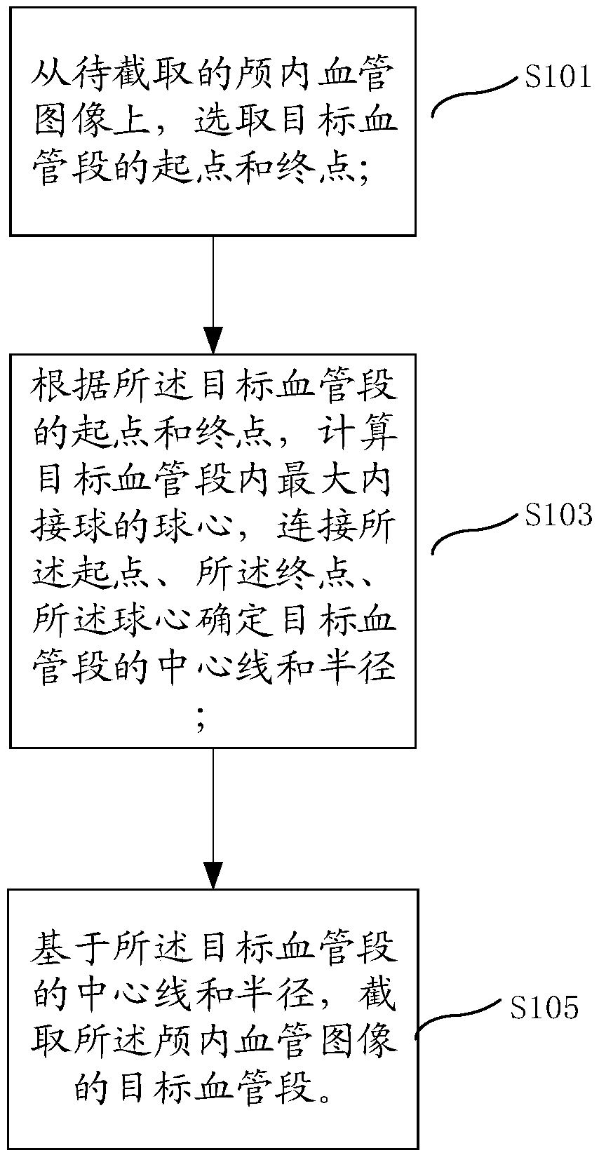
[0039] See figure 1 , This application provides a method for capturing images of intracranial blood vessels based on a centerline, including:

[0040] S101: Select the starting point and the end point of the target blood vessel segment from the intracranial blood vessel image to be intercepted;

[0041] S103: According to the start point and end point of the target blood vessel segment, calculate the sphere center of the largest inscribed ball in the target blood vessel segment, and connect the start point, the end point, and the sphere center to determine the center line and radius of the target blood vessel segment;

[0042] S105: Based on the centerline and radius of the target blood vessel segment, intercept the ta...


PUM


Abstract

Embodiments of the present application disclose a method and system for intercepting intracranial vascular images based on a centerline. The scheme includes selecting the starting point and the endingpoint of the target vessel segment from the intracranial vessel image to be intercepted; calculating a spherical center of the largest inward ball in the target blood vessel segment according to thestart point and the end point of the target blood vessel segment, connecting the start point and the end point, the spherical center determining a centerline and a radius of the target blood vessel segment; intercepting the target vessel segment of the intracranial vessel image based on the centerline and radius of the target vessel segment. This scheme realizes the local interception of the intracranial vascular image, which is convenient for the physicalization of intracranial artery, the simulation of interventional operation and the manufacture of related teaching equipment.

Description

Technical field [0001] This application relates to the field of medical imaging, in particular to a method and system for capturing images of intracranial blood vessels based on a centerline. Background technique [0002] With the continuous progress and development of modern medical imaging equipment, the development of medical imaging technology and computer graphics has brought tremendous changes to medicine. Computed tomography (CT), nuclear magnetic resonance (MRI), ultrasound (US), positron Transmitting computer tomography (PET), magnetic resonance angiography (magnetic resonance angiography, MRA), digital subtraction angiography (Digital Subtraction Angiography, DSA), CTA and other medical imaging technologies have been widely used in clinical diagnosis. [0003] In the prior art, three-dimensional images are used for diagnosis. Among them, the three-dimensional image includes: three-dimensional CTA image, three-dimensional MRA image, three-dimensional DSA image. [0004] Th...

Claims


Application Information

Patent Timeline
IPC IPC(8): G06T7/11
CPCG06T2207/10081G06T2207/10088G06T2207/30016G06T2207/30101G06T7/11
Inventor 杨光明冯雪王文智秦岚
Owner UNION STRONG (BEIJING) TECH CO LTD
Why Eureka
Try Eureka