Intracranial vascular image interception method and system based on a central line

A technology of intracranial blood vessels and central lines, applied in the field of medical imaging

Pending Publication Date: 2019-04-05
XUANWU HOSPITAL OF CAPITAL UNIV OF MEDICAL SCI
View PDF7 Cites 12 Cited by
  • Summary
  • Abstract
  • Description
  • Claims
  • Application Information

AI Technical Summary

Problems solved by technology

[0005] However, in the prior art, there is no local interception method for intracranial vascular images based on CTA images

Method used


Image

  • Intracranial vascular image interception method and system based on a central line
  • Intracranial vascular image interception method and system based on a central line
  • Intracranial vascular image interception method and system based on a central line

Examples


Embodiment Construction

[0037] Embodiments of the present application provide a centerline-based interception method and system for an intracranial vascular image, so as to solve the problem of partial interception of blood vessel segment images of the intracranial vascular image.

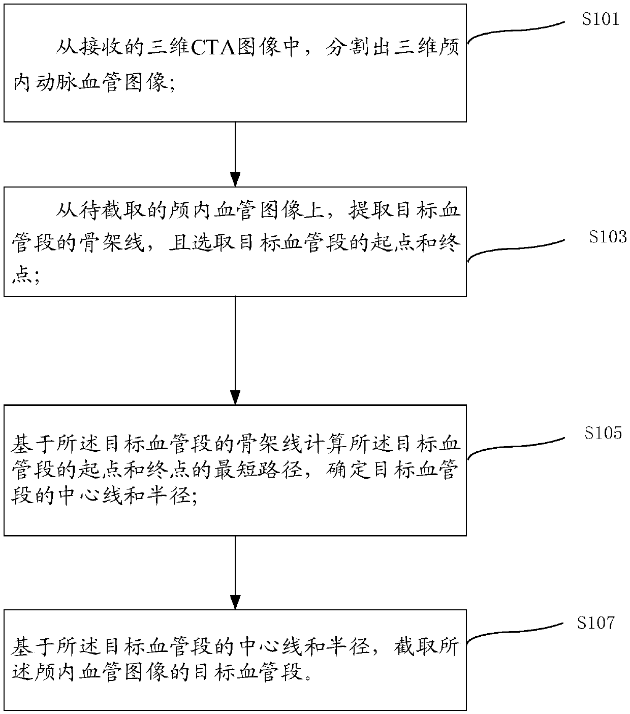
[0038] see figure 1 , the application provides a centerline-based interception method for intracranial blood vessel images, including:

[0039] S101: Segment a three-dimensional intracranial aneurysm vessel image from the received three-dimensional CTA image;

[0040] S103: From the intracranial blood vessel image to be intercepted, extract the skeleton line of the target blood vessel segment, and select the starting point and end point of the target blood vessel segment;

[0041] S105: Calculate the shortest path of the starting point and the end point of the target blood vessel segment based on the skeleton line of the target blood vessel segment, and determine the centerline and radius of the target blood vessel segme...


PUM


Abstract

The embodiment of the invention discloses an intracranial vascular image interception method and system based on a central line. The scheme comprises the steps of segmenting a three-dimensional intracranial artery blood vessel image from a received three-dimensional CTA image; Extracting a skeleton line of the target blood vessel segment from the intracranial blood vessel image to be intercepted,and selecting a starting point and a terminal point of the target blood vessel segment; Calculating the shortest path of the starting point and the ending point of the target blood vessel segment based on the skeleton line of the target blood vessel segment, and determining the center line and the radius of the target blood vessel segment; And based on the center line and the radius of the targetblood vessel segment, intercepting the target blood vessel segment of the intracranial blood vessel image. According to the scheme, the blood vessel segment image in the three-dimensional CTA image islocally captured, so that the intracranial artery blood vessel segment can be materialized, and interventional operation simulation and related teaching appliance manufacturing are facilitated.

Description

technical field [0001] The present application relates to the field of medical imaging, in particular to a centerline-based interception method and system for intracranial blood vessel images. Background technique [0002] With the continuous progress and development of modern medical imaging equipment, the development of medical imaging technology and computer graphics has brought great changes to medicine. Computed tomography (CT), nuclear magnetic resonance (MRI), ultrasound (US), positron Emission computed tomography (PET), magnetic resonance angiography (magnetic resonance angiography, MRA), digital subtraction angiography (Digital Subtraction Angiography, DSA), CTA and other medical imaging techniques have been widely used in clinical diagnosis. [0003] In the prior art, a three-dimensional CTA image is a common image for image diagnosis. [0004] CTA is an angiographic examination using CT (Computer-Aided Tomography) technology, which can detect all arteries and vei...

Claims


Application Information

Patent Timeline
Patent Type & Authority Applications(China)
IPC IPC(8): G06T5/00G06T7/10
CPCG06T5/00G06T7/10
Inventor 叶明胡鹏何川孙力泳杨光明冯雪王文智秦岚
Owner XUANWU HOSPITAL OF CAPITAL UNIV OF MEDICAL SCI
Why Eureka
Try Eureka