
CT image abdominal multi-organ segmentation method and device

A CT image, multi-organ technology, applied in the field of image processing, can solve the problem of poor accuracy of segmentation of multiple organs in the abdomen of three-dimensional CT images, and achieve the effect of stable segmentation results

Inactive Publication Date: 2019-09-10
BEIJING INSTITUTE OF TECHNOLOGYGY
View PDF7 Cites 11 Cited by
  • Summary
  • Abstract
  • Description
  • Claims
  • Application Information

AI Technical Summary

Problems solved by technology

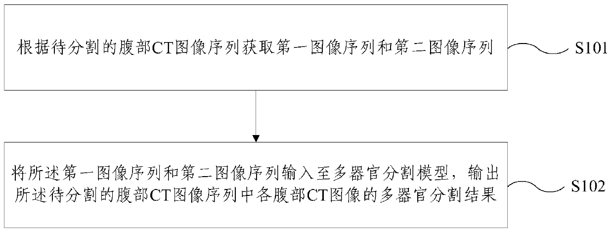
[0005] Embodiments of the present invention provide a method and device for segmenting multiple organs in the abdomen in CT images to solve or at least partially solve the defect of poor accuracy in the segmentation of multiple organs in the abdomen in 3D CT images in the prior art

Method used


Image

  • CT image abdominal multi-organ segmentation method and device
  • CT image abdominal multi-organ segmentation method and device
  • CT image abdominal multi-organ segmentation method and device

Examples


Embodiment Construction

[0039] In order to make the purpose, technical solutions and advantages of the embodiments of the present invention clearer, the technical solutions in the embodiments of the present invention will be clearly and completely described below in conjunction with the drawings in the embodiments of the present invention. Obviously, the described embodiments It is a part of embodiments of the present invention, but not all embodiments. Based on the embodiments of the present invention, all other embodiments obtained by persons of ordinary skill in the art without creative efforts fall within the protection scope of the present invention.

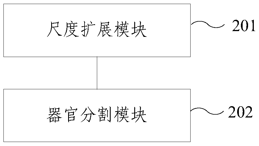
[0040] In order to overcome the above-mentioned problems in the prior art, an embodiment of the present invention provides a method and device for segmenting multiple organs in the abdomen of a CT image. information, so as to obtain more accurate and stable segmentation results.

[0041] figure 1 It is a schematic flowchart of a method for segme...


PUM


Abstract

The embodiment of the invention provides a CT image abdominal multi-organ segmentation method and device. The method comprises the steps of obtaining a first image sequence and a second image sequenceaccording to an abdominal CT image sequence to be segmented; inputting the first image sequence and the second image sequence into a multi-organ segmentation model, and outputting a multi-organ segmentation result of each abdominal CT image in the abdominal CT image sequence to be segmented; wherein the scale of the image in the first image sequence is greater than that of the image in the secondimage sequence; wherein the multi-organ segmentation model is obtained after training based on abdominal CT image sequence sample data and a predetermined manual segmentation marking result. According to the CT image abdominal multi-organ segmentation method and device provided by the embodiment of the invention, the multi-organ segmentation result of each abdominal CT image in the abdominal CT image sequence to be segmented is obtained according to the context information of the organ levels obtained by the two different scales, and a more accurate and stable segmentation result can be obtained.

Description

technical field [0001] The present invention relates to the technical field of image processing, and more particularly, to a method and device for segmenting abdominal multiple organs in a CT image. Background technique [0002] Organ segmentation refers to determining the regions belonging to each organ in medical images and identifying organs in medical images. Segmentation of multiple abdominal organs in CT images can identify the abdominal organs in medical images. There are many vital organs in the human abdominal cavity, including the liver, gallbladder, pancreas, spleen and kidneys. Multi-organ segmentation refers to segmenting the region included in each target organ in the image, and the number of target organs is multiple. The contrast of abdominal CT images is low, and the boundaries of various organs in the images are blurred and various. Affected by abdominal pressure, some organs in the human body (such as stomach, pancreas, liver, etc.) have large deformatio...

Claims


Application Information

Patent Timeline
Patent Type & Authority Applications(China)
IPC IPC(8): G06T7/10
CPCG06T7/10G06T2207/10081G06T2207/10016G06T2207/20084G06T2207/20081
Inventor 艾丹妮杨健王涌天范敬凡谢桂望
Owner BEIJING INSTITUTE OF TECHNOLOGYGY
Learn More