PET-CT lung tumor segmentation method combining three dimensional graph cut algorithm with random walk algorithm

A random walk and graph cut algorithm technology, applied in the field of biomedical image processing, can solve the problems of low segmentation accuracy and reliability, and cannot bring qualitative changes to clinical applications.

Active Publication Date: 2016-06-22
SUZHOU BIGVISION MEDICAL TECH CO LTD
View PDF4 Cites 23 Cited by
  • Summary
  • Abstract
  • Description
  • Claims
  • Application Information

AI Technical Summary

Problems solved by technology

[0003] However, many existing lung tumor methods at home and abroad are based on a single modality (PET or CT), such as: region growing method, LevelSet method, etc., the accuracy and reliability of the segmentation are not very high, and it cannot really be used in clinical practice. Application brings about qualitative change

Method used


Image

  • PET-CT lung tumor segmentation method combining three dimensional graph cut algorithm with random walk algorithm
  • PET-CT lung tumor segmentation method combining three dimensional graph cut algorithm with random walk algorithm
  • PET-CT lung tumor segmentation method combining three dimensional graph cut algorithm with random walk algorithm

Examples


Embodiment Construction

[0045] The present invention will be further described below in conjunction with the accompanying drawings.

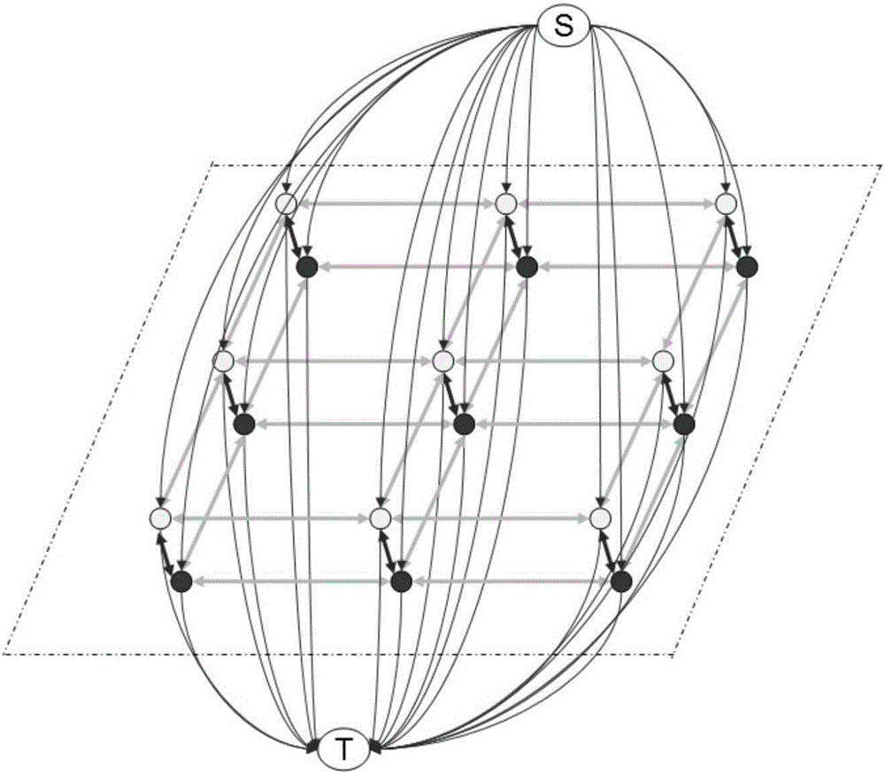
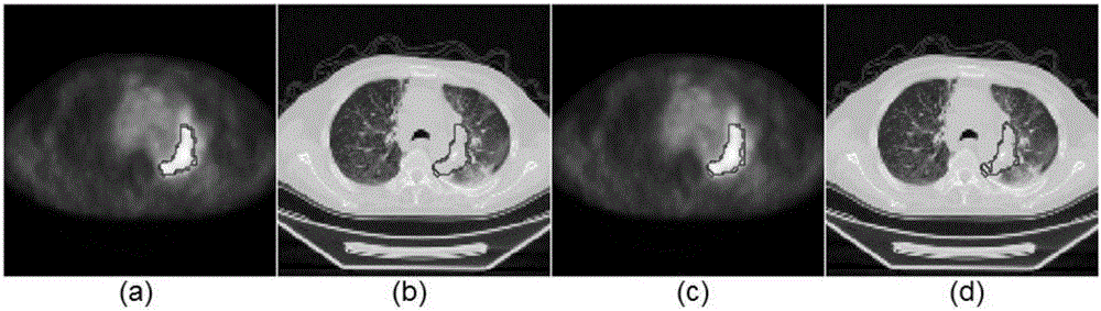
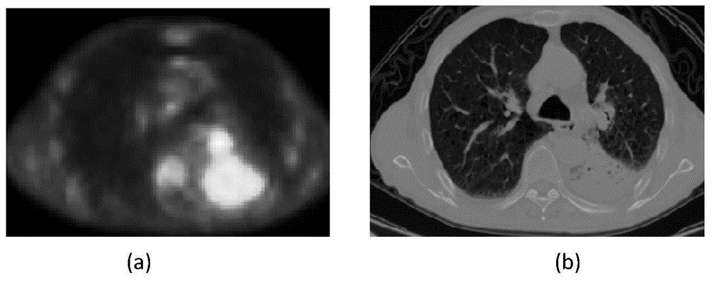
[0046] The lung tumor segmentation method of the present invention is based on the joint segmentation of the two modalities of PET-CT, and is intended to make full and reasonable use of the feature information of the two modalities for precise positioning and segmentation. Such as figure 2 It is a slice image of lung PET and CT. The specific division method is as follows:

[0047] 1. Carry out a linear upsampling operation on the original PET image, so that the PET image and the CT image have the same pixel points, and perform affine registration on the PET and CT images, and finally make the pixels of the PET and CT images correspond one-to-one. And judge whether it belongs to tumor or non-tumor area by human eyes, manually calibrate the tumor seed point and non-tumor seed point of the image, and at the same time determine the gold standard of lung tumor under the ...


PUM


Abstract

The invention belongs to the field of biomedical image processing and specifically relates to a PET-CT lung tumor segmentation method combining a three dimensional graph cut algorithm with a random walk algorithm. The method comprises the following steps of: performing linear up-sampling on an original PET image and performing affine registration on PET and CT images; calibrating tumor seed points and non-tumor seed point; performing random walk algorithm segmentation on the PET image in combination with the tumor seed points; acquiring a foreground target area Ro completely including a target lung tumor area, using the areas, except the Ro, as a background area Rb of a non-lung tumor area; establishing gauss mixture models for the foreground area Ro and the background area Rb separately; computing energy items according to the gauss mixture models of the foreground area Ro and the background area Rb and obtaining a final segmentation result by using an graph cut algorithm. The method fully utilizes the function information and the PET image and the structure information of the CT image, enables complements between the random walk algorithm and the graph cut algorithm, and achieves an accurate lung tumor segmentation result.

Description

technical field [0001] The invention belongs to the field of biomedical image processing, and utilizes a Gaussian Mixture Models (Gaussian MixtureModels) optimized Graph Cut (GraphCut) method combined with a random walk (Randomwalk) algorithm to accurately segment lung tumors. Background technique [0002] Most lung tumors originate from the bronchial mucosal epithelium, so it is also called bronchial lung cancer. In the past 50 years, the incidence of lung cancer has increased significantly all over the world. According to statistics, in some countries in Europe and the United States and in large cities in my country, the incidence of lung cancer has ranked first among all male tumors. The treatment of lung tumors requires tumor location, size and shape analysis, and PET (Positron Emission Computed Tomography) and CT (Computed Tomography), as two quantitative molecular-structural imaging techniques, have been widely used in the analysis and diagnosis of lung tumors. PET ima...

Claims


Application Information

Patent Timeline
Patent Type & Authority Applications(China)
IPC IPC(8): G06T7/00
CPCG06T7/0012G06T2207/10012G06T2207/10081G06T2207/10104G06T2207/30061G06T2207/30096
Inventor 陈新建俞凯向德辉朱伟芳石霏
Owner SUZHOU BIGVISION MEDICAL TECH CO LTD
Why Eureka
Try Eureka