Segmentation method and system for abdomen soft tissue nuclear magnetism image

A technology of nuclear magnetic image and soft tissue, which is applied to the field of organ tissue segmentation algorithm of abdominal nuclear magnetic image, and can solve the problem of low computational efficiency of the algorithm.

Active Publication Date: 2013-12-25
SHENZHEN INST OF ADVANCED TECH CHINESE ACAD OF SCI
View PDF3 Cites 23 Cited by
  • Summary
  • Abstract
  • Description
  • Claims
  • Application Information

AI Technical Summary

Problems solved by technology

The patent application number 201210123996 proposes an automatic segmentation and realization method of adaptive external force level set of soft tissue MRI images, but requires manual interaction to select the initial contour, iterative execution makes the calculation efficiency of the algorithm not high, and has certain limitations in application

Method used


Image

  • Segmentation method and system for abdomen soft tissue nuclear magnetism image
  • Segmentation method and system for abdomen soft tissue nuclear magnetism image
  • Segmentation method and system for abdomen soft tissue nuclear magnetism image

Examples


Embodiment 1

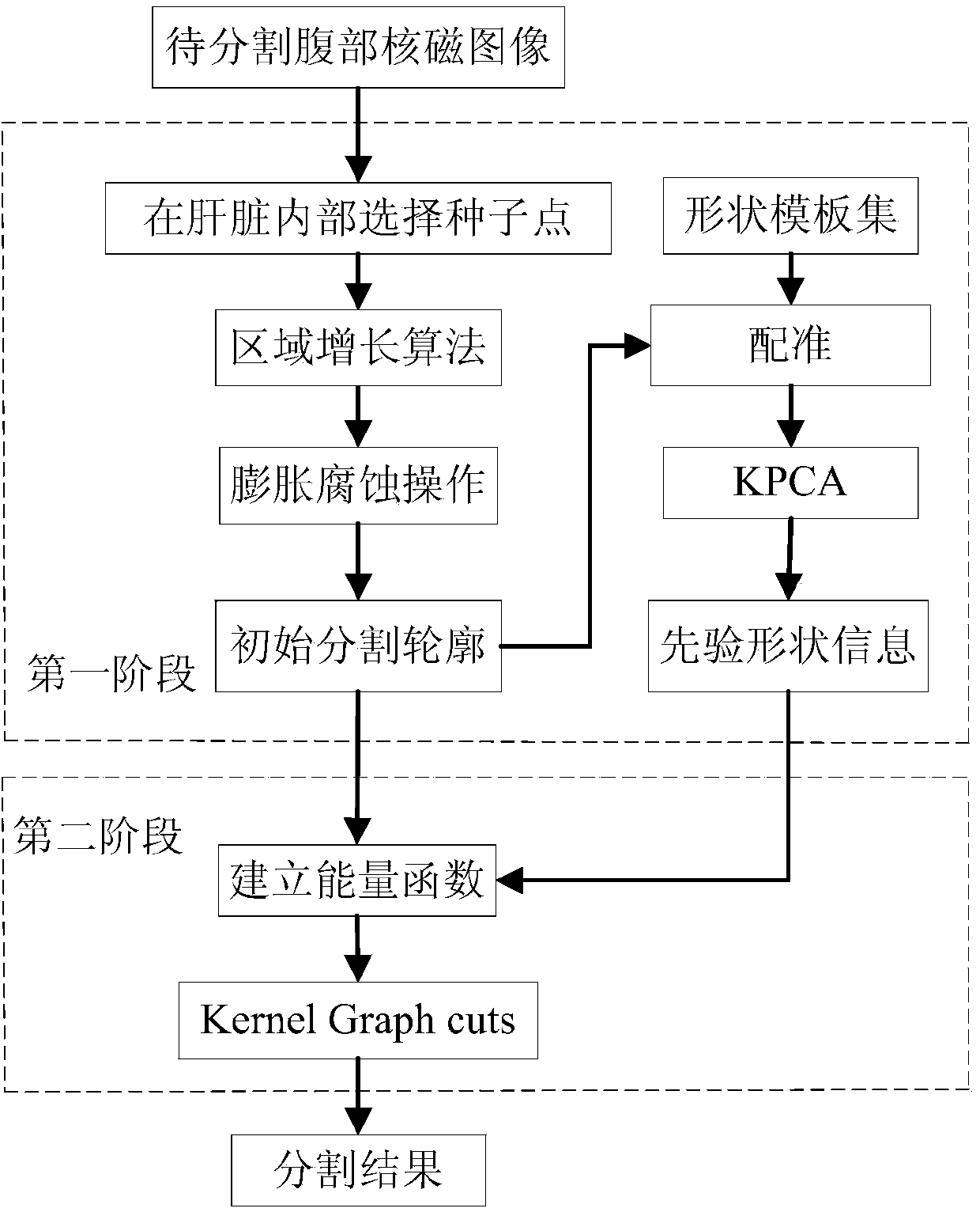
[0056] A method for segmenting liver tissue using abdominal soft tissue nuclear magnetic image segmentation, such as figure 1 shown, including the following steps:

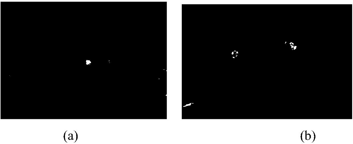
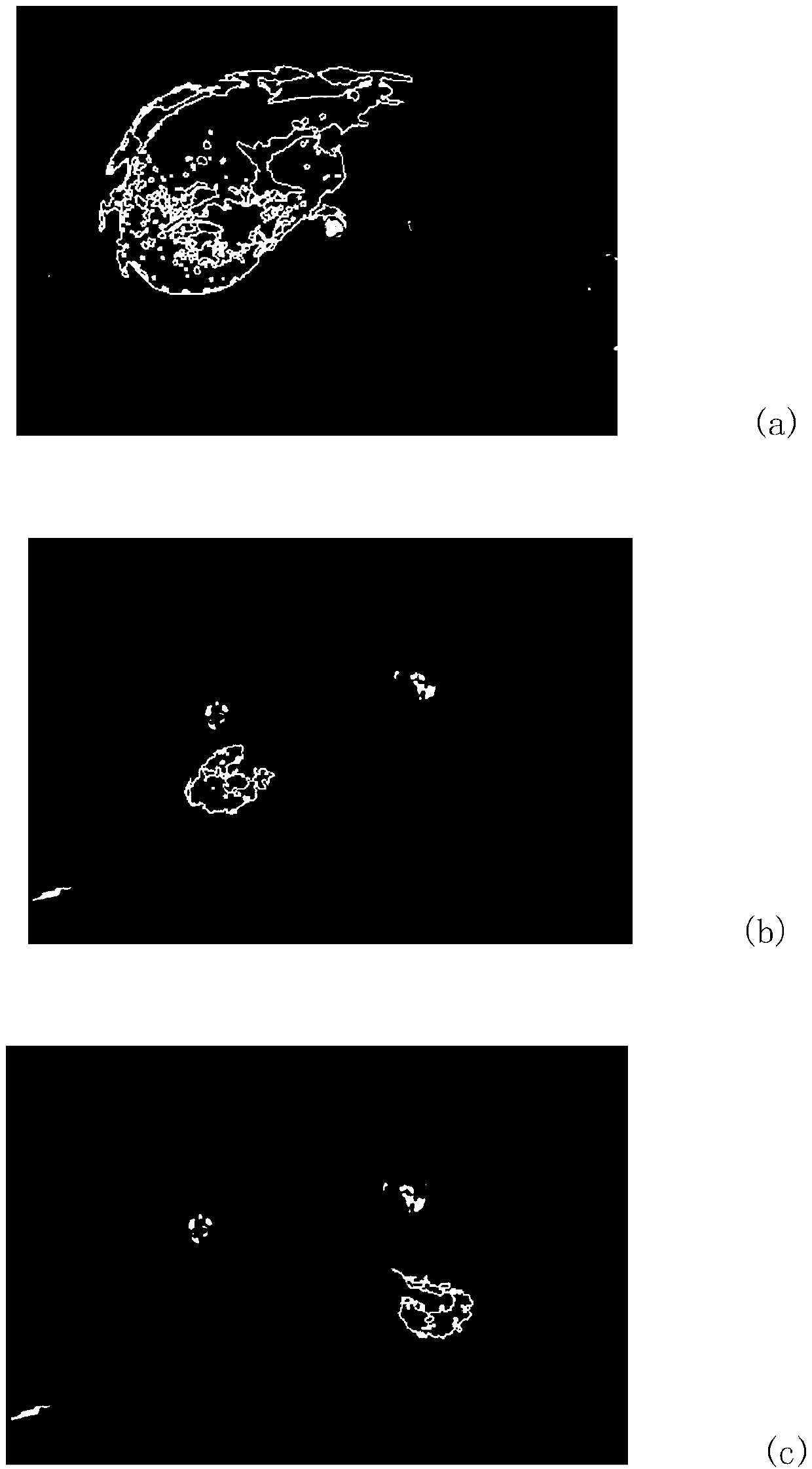
[0057] (1) Select a seed point inside the liver. The seed point is selected manually, mainly within the segmented region. The region growing algorithm is used to perform region growth with the selected seed point as the initial point, and the pre-segmentation is performed inside the liver to obtain the predicted value. Segmentation area; liver MRI image to be segmented such as figure 2 As shown in (a), the pre-segmentation results are as follows image 3 as shown in (a); from image 3 In (a), it can be seen that there are many isolated areas inside the liver that have not been segmented.

[0058] (2) Dilate and corrode the pre-segmented area by using morphological operators to form an initial segmentation contour inside and outside the pre-segmented area; both dilation and erosion operations use the same size ...

Embodiment 2

[0086] The method for segmenting kidney tissue using the method for segmenting abdominal soft tissue MRI images comprises the following steps:

[0087] (1) Select a seed point inside the kidney. The seed point is selected manually, mainly within the segmented region. The region growing algorithm is used to perform region growth with the selected seed point as the initial point, and the pre-segmentation is performed inside the kidney to obtain the predicted value. Segmentation area; kidney MRI image to be segmented such as figure 2 As shown in (b), the pre-segmentation results are as follows image 3 in (b) and (c); from image 3 In (b) and (c), it can be seen that there are many isolated areas inside the kidney that have not been segmented.

[0088] (2) Dilate and corrode the pre-segmented area by using morphological operators to form an initial segmentation contour inside and outside the pre-segmented area; both dilation and erosion operations use the same size structural ...


PUM


Abstract

The invention discloses a segmentation method and system for an abdomen soft tissue nuclear magnetism image. The segmentation method comprises the steps that pre-segmentation is conducted on an area to be segmented through an area growing algorithm, then a morphological operator is adopted to conduct expansion and corrosion operations to carry out further processing on the pre-segmentation result, so that the pre-segmentation result forms an original segmentation outline. After rectification is conducted between a shape template set and the original segmentation outline, kernel principal component analysis is conducted, and prior shape information is obtained through a statistics model. The prior shape information is combined with data items of an energy function of a nuclear magnetism image segmentation model, and an energy function is built; a kernel graph cuts algorithm is used for carrying out segmentation on the original segmentation outline and an objective outline is obtained. The segmentation method and system can achieve semi-automatic segmentation, the system is simple, the robustness of the nuclear magnetism image segmentation algorithm is effectively improved so as to enable the segmentation result to be more accurate, and the segmentation method and system for the abdomen soft tissue nuclear magnetism image can be applied to nuclear magnetism image segmentation.

Description

technical field [0001] The invention relates to the field of organ tissue segmentation algorithms for abdominal nuclear magnetic images, in particular to a method and system for abdominal soft tissue nuclear magnetic image segmentation. Background technique [0002] Magnetic resonance imaging (magnetic resonance imaging, MRI) uses radio frequency (radio frequency, RF) electromagnetic waves to excite substances containing atomic nuclei with non-zero spins in a magnetic field, and nuclear magnetic resonance (nuclear magnetic resonance, NMR) occurs. A digital image is established by collecting magnetic resonance signals with induction coils and processing them according to certain mathematical methods. The NMR signal intensity is related to the density of hydrogen nuclei in the sample. Two-thirds of the weight of the human body is water, and different tissues have different proportions of water molecules, so the NMR signal intensity is also different. This difference is used as...

Claims


Application Information

Patent Timeline
Patent Type & Authority Applications(China)
IPC IPC(8): G06T7/00
Inventor 秦文健罗清辜嘉
Owner SHENZHEN INST OF ADVANCED TECH CHINESE ACAD OF SCI
Why Eureka
Try Eureka