Retinal macular edema multi-lesion image segmentation method

A macular edema and image segmentation technology, applied in the field of image processing, can solve the problem of simultaneous segmentation of multi-lesion images of retinal macular edema and multiple lesions, so as to solve the problem of huge data imbalance, improve the segmentation performance, and alleviate the huge data imbalance. balanced effect

Active Publication Date: 2019-10-18
SUZHOU UNIV
View PDF8 Cites 16 Cited by
  • Summary
  • Abstract
  • Description
  • Claims
  • Application Information

AI Technical Summary

Problems solved by technology

[0004] The purpose of the present invention is to overcome the deficiencies in the prior art, provide a kind of retinal macular edema multi-lesion image segmenta

Method used


Image

  • Retinal macular edema multi-lesion image segmentation method
  • Retinal macular edema multi-lesion image segmentation method
  • Retinal macular edema multi-lesion image segmentation method

Examples


Embodiment Construction

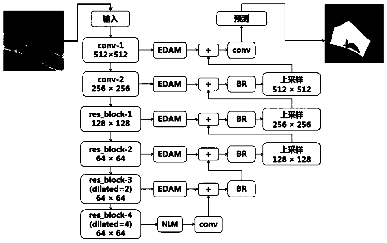
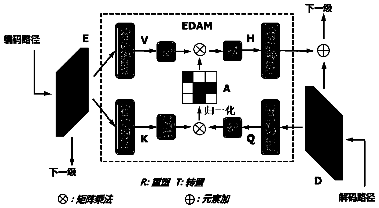
[0057] The embodiment of the present invention provides a method for image segmentation of retinal macular edema with multiple lesions. The pre-built and trained codec attention network model is used to realize the joint segmentation of retinal macular edema and multiple lesions, and the results of segmented images can be obtained, which can predict macular edema. Multiple lesions in multi-lesion images are segmented simultaneously, laying the foundation for subsequent quantitative analysis of lesions. The present invention will be further described below in conjunction with the accompanying drawings from the design and construction of image preprocessing, network model and experimental comparison results. The following examples are only used to more clearly illustrate the technical scheme of the present invention, but cannot limit the protection scope of the present invention with this.

[0058] 1. Image preprocessing:

[0059] In order to prevent GPU memory overflow, the th...


PUM


Abstract

The invention discloses a retinal macular edema multi-lesion image segmentation method. The method comprises the steps of collecting three-dimensional retinal OCT volume data and converting the three-dimensional retinal OCT volume data into a two-dimensional retinal OCT B scanning image; and inputting the two-dimensional retina OCT B scanning image into a trained coding and decoding attention network model to carry out retina macular edema multi-lesion joint segmentation, and obtaining a segmentation image result. According to the method, the coding and decoding attention network model is adopted for multi-lesion joint segmentation, richer global features can be obtained, simultaneous segmentation of retinal edema, subretinal effusion and pigment epithelium detachment multi-lesion in the retinal macular edema multi-lesion image is achieved, and a foundation is laid for follow-up lesion quantitative analysis.

Description

technical field [0001] The invention relates to a method for segmenting images of retinal macular edema with multiple lesions, and belongs to the technical field of image processing. Background technique [0002] According to statistics, about 34% of diabetic patients will develop diabetic retinopathy (referred to as sugar net, Diabet Retinopathy, DR). Diabetic reticulum is a common blinding disease. Severe diabetic reticulum disease is often accompanied by retinal edema (Retinal Edema, RE), sub-retinal fluid (Sub-Retinal Fluid, SRF) and pigment epithelial detachment (Pigment epithelial detachment). Epithelial Detachment, PED) and other symptoms. The quantitative segmentation of retinal edema, subretinal fluid and pigment epithelial detachment can provide important reference information for clinical analysis and research of diabetic reticulum. However, simultaneous segmentation of multiple lesion regions in Optical Coherence Tomography (OCT) images is very challenging, mai...

Claims


Application Information

Patent Timeline
IPC IPC(8): G06T7/10G06T7/11G06T3/00
CPCG06T3/0037G06T2207/10012G06T2207/10101G06T2207/20081G06T2207/30041G06T2207/30096G06T7/10G06T7/11
Inventor 朱伟芳冯爽朗陈新建赵鹤鸣
Owner SUZHOU UNIV
Why Eureka
Try Eureka