Brain slice image region division method and device

An image area and brain slice technology, applied in the field of image registration, can solve the problems of inaccurate area division method and inaccurate area division device division, and achieve the effects of rapid area division, accurate area division, and simple and convenient operation.

Active Publication Date: 2019-12-10
ZHENGZHOU UNIV
View PDF4 Cites 8 Cited by
  • Summary
  • Abstract
  • Description
  • Claims
  • Application Information

AI Technical Summary

Problems solved by technology

[0004] The purpose of the present invention is to provide a brain slice image region division method to solve the problem of inaccurate division of existing region division methods; meanwhile, it also provides a brain slice image region division device to solve the problem of division of existing region division devices. inaccurate question

Method used


Image

  • Brain slice image region division method and device
  • Brain slice image region division method and device
  • Brain slice image region division method and device

Examples


Embodiment Construction

[0037] Brain slice image region division method embodiment:

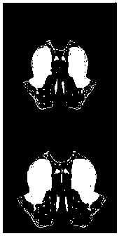
[0038] The brain slice image area division method proposed in this embodiment, such as figure 1 shown, including the following steps:

[0039] 1) Affine transformation, pre-registration.

[0040] The purpose of this step is to obtain multiple groups of images, each group of images includes the original image to be divided (here is the brain slice image) and the corresponding standard brain atlas image, and the standard brain atlas image is affine transformed to realize brain Pre-registration of atlas images.

[0041] The brain slice images here are brain slice images of immunohistochemistry and confocal imaging with a size of m*m, and the corresponding standard brain atlas images are two kinds of brain atlas images: Average Template brain atlas and Atlas brain atlas. Moreover, the standard brain atlas image includes an Average Template brain atlas and an Atlas brain atlas, the Average Template brain atlas is a gr...


PUM


Abstract

The invention relates to a brain slice image region division method and device, and belongs to the technical field of image registration. The method comprises the steps of acquiring multiple groups ofimages wherein each group of images comprises an original image to be subjected to region division and a gray scale and color standard brain atlas corresponding to the original image; performing affine transformation on the gray scale and color standard brain atlas to obtain a gray scale and color standard brain atlas after affine transformation; respectively carrying out feature extraction on the original image and the affine-transformed gray scale standard brain atlas to obtain a single-mode original image and a single-mode gray scale standard brain atlas; taking the single-mode original image and the single-mode gray scale standard brain atlas as training samples, and training a registration network; inputting the color standard brain map after affine transformation into a trained registration network to obtain a registered color standard brain map; and fusing with an original image to complete region division. Full-automatic and rapid region division of the multi-modal brain sliceimage is realized, the operation is simple and convenient, and the region division is accurate.

Description

technical field [0001] The invention relates to a brain slice image region division method and device, belonging to the technical field of image registration. Background technique [0002] The brain is the most complex organ in the animal body. It is composed of a large number of cells with different shapes and functions, and controls various psychological and physiological activities of animals through different neural networks. Studying the brain is the basis for treating neurological diseases, simulating brain network functions, and optimizing artificial intelligence. The emergence and extensive application of neural tissue staining methods such as immunohistochemical staining, acetylcholinesterase staining, and Nissl staining have prompted people to produce standard brain atlases with fine brain structures in various animals such as mice, rats, and humans. With the further development of neural circuit labeling and neural staining technology and the application of magne...

Claims


Application Information

Patent Timeline
Patent Type & Authority Applications(China)
IPC IPC(8): G06T7/00G06T7/11G06T7/13G06T7/33
CPCG06T7/0014G06T7/11G06T7/13G06T7/337G06T2207/20081G06T2207/20221G06T2207/30016
Inventor 王松伟赵秋阳师丽
Owner ZHENGZHOU UNIV
Why Eureka
Try Eureka