Eureka AIR delivers breakthrough ideas for toughest innovation challenges, trusted by R&D personnel around the world.

Medical image processing method and device

A technology of medical imaging and processing method, applied in the field of computer, can solve the problems of large influence of subjective factors of mapping accuracy and low efficiency, etc.

Active Publication Date: 2020-01-17
HANGZHOU YITU MEDIAL TECH CO LTD
View PDF7 Cites 5 Cited by
  • Summary
  • Abstract
  • Description
  • Claims
  • Application Information

AI Technical Summary

Problems solved by technology

[0005] The embodiment of the present invention provides a medical image processing method and device to solve the technical problems in the prior art that the mapping accuracy is greatly affected by human subjective factors and the efficiency is low due to manual mapping of 2D medical images and panoramas

Method used


Image

  • Medical image processing method and device
  • Medical image processing method and device
  • Medical image processing method and device

Examples


Embodiment Construction

[0038] In order to make the purpose, technical solutions and advantages of the present invention clearer, the present invention will be further described in detail below in conjunction with the accompanying drawings. Obviously, the described embodiments are only some of the embodiments of the present invention, rather than all of them. Based on the embodiments of the present invention, all other embodiments obtained by persons of ordinary skill in the art without making creative efforts belong to the protection scope of the present invention.

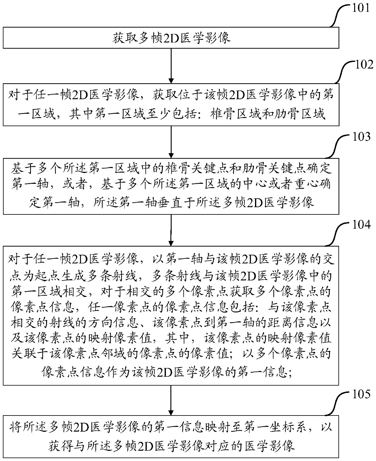
[0039] The medical image processing method in the embodiment of the present invention can map multiple frames of 2D medical images, for example, it can map multiple frames of cross-sectional images of the chest taken by CT equipment to images including complete vertebrae and ribs (ie panoramic images) , and the corresponding relationship with multiple frames of 2D medical images can be marked in the mapped panorama. It should be noted t...


PUM


Abstract

The embodiment of the invention discloses a medical image processing method and device. The method comprises the steps of: determining a first area in each frame of 2D medical image, determining a first axis according to the first areas, generating a plurality of rays by taking the intersection point of the first axis and each frame of 2D medical image as a starting point, determining pixel pointinformation of the plurality of pixel points where the plurality of rays intersect with the first areas in each frame of 2D medical image, taking the pixel point information as first information of each frame of 2D medical image, and mapping the first information of the plurality of frames of 2D medical images to a first coordinate system to obtain medical images corresponding to the plurality offrames of 2D medical images. The first information of each frame of 2D medical image is determined in a ray radiation mode and is mapped to the first coordinate system to automatically map the multi-frame 2D medical images to a panorama with no need for artificial and subjective determination of a corresponding relationship of the 2D medical images and the panorama, and therefore, the efficiency and the precision of mapping are improved.

Description

technical field [0001] The invention relates to the field of computer technology, in particular to a medical image processing method and device. Background technique [0002] With the development of modern medical imaging technology, various medical imaging devices are constantly emerging, such as X-ray photography equipment, computed tomography (Computed Tomography, CT) equipment, nuclear magnetic resonance imaging (Nuclear Magnetic Resonance Imaging, NMRI) equipment and so on. These medical imaging devices can collect cross-sectional images (also called 2D images) of various parts of the human body, and can also generate 3D images of various parts of the human body. [0003] At this stage, the cross-sectional images collected by medical imaging equipment usually cannot correspond to the panorama, so manual judgment is required to match the tissue in the cross-sectional image with the tissue in the panorama. Taking the collected chest image as an example, the chest image i...

Claims


Application Information

Patent Timeline
Patent Type & Authority Applications(China)
IPC IPC(8): G16H30/00
CPCG16H30/00
Inventor 倪浩石磊魏子昆华铱炜柏慧屏
Owner HANGZHOU YITU MEDIAL TECH CO LTD
Why Eureka
Eureka Blog
Learn More