A fluorescent probe that uses fluorescent images to display two states of cell membrane potential and its application
A fluorescent probe and fluorescent image technology, applied in the field of fluorescent probes, to achieve high specificity and good application prospects
- Summary
- Abstract
- Description
- Claims
- Application Information
AI Technical Summary
Problems solved by technology
Method used
Image

Examples
Embodiment 1
[0029] Example 1 Preparation of the Fluorescent Probe (CP12) Using Fluorescent Images to Display Two States of Cell Membrane Potential in the Present Invention
[0030] Add 0.399g of N-dodecylcarbazole-3-carbaldehyde and 5ml of ethanol to a dry three-necked flask, protect it with nitrogen, heat up to 60°C, and dissolve it after heating for 30min. Add the compound 1-(2-hydroxyethyl )-4-picoline iodide salt 0.265g, add 5 drops of piperidine dropwise, heat up to reflux, stop heating overnight, evaporate the solvent after cooling, recrystallize with ethanol, dry to obtain 0.2g of yellow-green powder, the yield 51%, which is CP12.
[0031] Above-mentioned compound CP12 preparation reaction formula is as follows:
[0032]
[0033] 1 1H NMR (300MHz, DMSO-d6), δ (ppm): 8.83 (d, J = 6.9Hz, 2H), 8.60 (d, J = 0.9Hz, 1H), 8.19-8.26 (m, 4H), 7.90 ( d, J=9Hz, 1H), 7.74(d, J=8.7Hz, 1H), 7.67(d, J=8.4Hz, 1H), 7.49-7.58(m, 2H), 7.29(t, J=7.5Hz ,1H),5.27(t,J=5.4Hz,1H),4.54(t,J=4.5Hz,2H),4....
Embodiment 2
[0034] Embodiment 2 cancer cell (SiHa and HeLa) and normal cell (Fibroblast) culture
[0035] at 37°C, 5% CO 2 In a saturated humidity incubator, SiHa, HeLa and Fibroblast cell lines were grown adherently in H-DMEM medium containing 10% fetal bovine serum (containing 1% double antibody). After the cells grew to the logarithmic phase, the SiHa, HeLa, and Fibroblast cells were taken and cultured. The specific method is: ① Soak the coverslip in absolute ethanol for 30 minutes, dry it with an alcohol lamp, and put it into a disposable 35mm culture dish; ② Wash the cells that have grown to logarithmic phase in the cell dish three times with PBS, After digesting with 1mL 0.25% trypsin for 3-5 minutes, pour out the enzyme solution carefully, then add a small amount of fresh culture medium, pipette the cells to make a uniform cell suspension, after counting the cells, use the cell culture medium to adjust the final concentration of the cells 1×10 5 cells / ml, inoculated into a petri...
Embodiment 3
[0036] Example 3 Fluorescent probe CP12 stains living SiHa, HeLa and Fibroblast cells
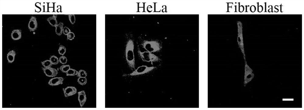
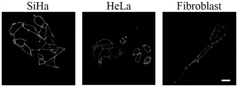
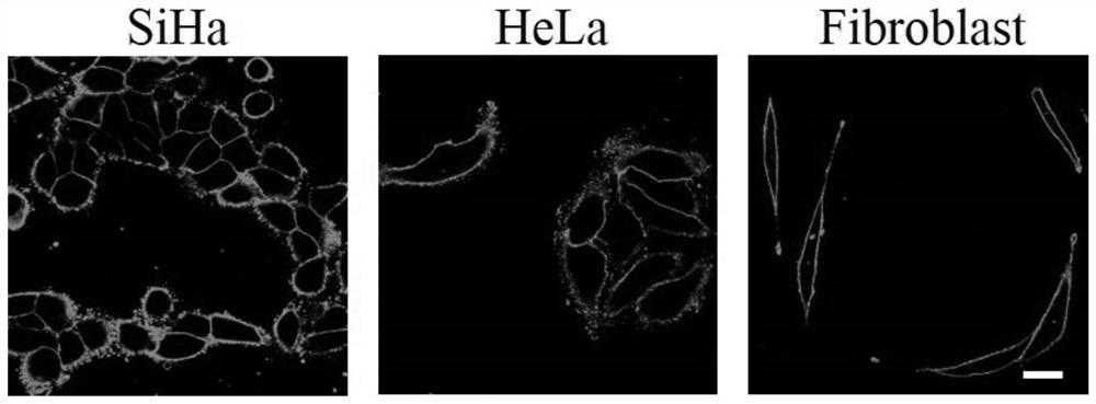
[0037] Wash the cell slides prepared in Example 2 three times with PBS, stain live SiHa, HeLa and Fibroblast cells with 2 μM CP12 respectively, incubate at room temperature for 20 min, suck out the culture medium, and wash three times with PBS to wash away unbound cells. The excess staining solution, the cell growth side was covered on the glass slide, under the excitation of 473nm laser, the laser scanning confocal fluorescence microscope was used to observe the coloring part of the cell, the fluorescence distribution and the change of brightness, etc. It was found that the fluorescent probe CP12 can pass through The cell membrane enters the cell, and both the cell membrane and the cytoplasm are stained with bright green fluorescence.
[0038] see results figure 1 .
[0039] The results indicated that the membrane potential state of the living cells was normal. When the cells were in the...
PUM
 Abstract
Description
Claims
Application Information
 - R&D Engineer
- R&D Manager
- IP Professional
- Industry Leading Data Capabilities
- Powerful AI technology
- Patent DNA Extraction
Browse by: Latest US Patents, China's latest patents, Technical Efficacy Thesaurus, Application Domain, Technology Topic.
© 2024 PatSnap. All rights reserved.Legal|Privacy policy|Modern Slavery Act Transparency Statement|Sitemap