
Opthalmoscope using natural pupil dilation

A technology of pupils and lenses, applied in the field of medical diagnostic devices

Active Publication Date: 2020-05-05
IBM CORP
View PDF3 Cites 3 Cited by
  • Summary
  • Abstract
  • Description
  • Claims
  • Application Information

AI Technical Summary

Problems solved by technology

The increased FOV allows medical professionals to examine the internal anatomy of the eye. In the prior art, it was thought that visual inspection using white light without full pupil dilation, and the use of various devices and imaging techniques produced little to no beneficial outcome

Method used


Image

  • Opthalmoscope using natural pupil dilation
  • Opthalmoscope using natural pupil dilation
  • Opthalmoscope using natural pupil dilation

Examples


Embodiment Construction

[0044] Current ophthalmoscope devices and procedures have several major medical and practical shortcomings. Patients and medical professionals must endure the disadvantages of these practices, it is a fact in life, and that millions of people around the world simply cannot get conventional treatments have been accepted. This is especially the case because there are no known alternatives other than to completely abandon the inspection or perform other inspections with limited benefits and incomplete results.

[0045] The embodiments of the present invention solve at least four types of problems in this field: (1) pupil dilation induction, (2) sequential inspection, (3) synchronization and (4) mobility and scale.

[0046] Pupil dilation is induced. Chemically induced pupil dilation can cause long-term discomfort. The patient's eyes remain dilated for much longer than the time of using the ophthalmoscope. When the pupils of the patient are dilated, the patient will experience blurr...


PUM


Abstract

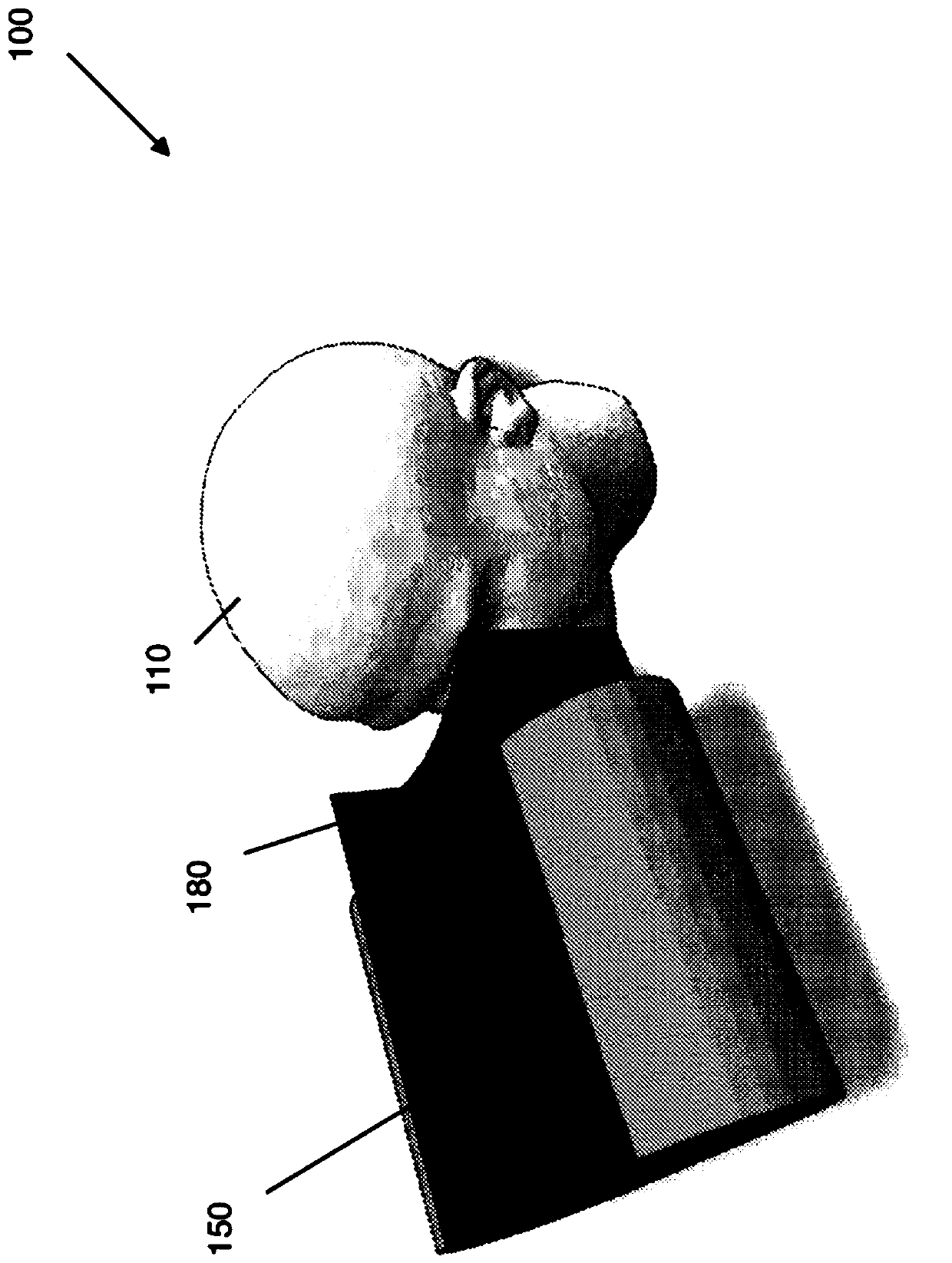
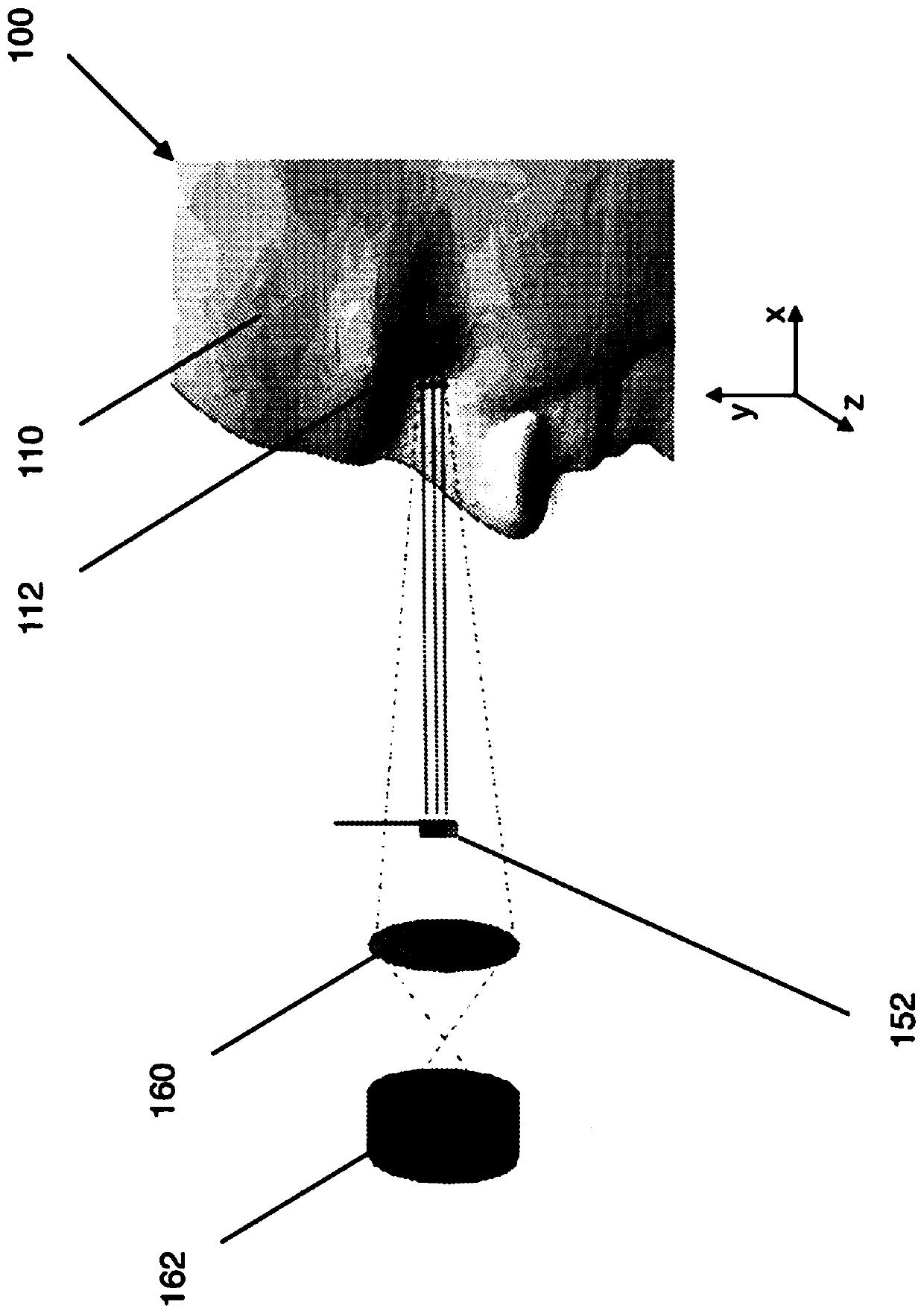
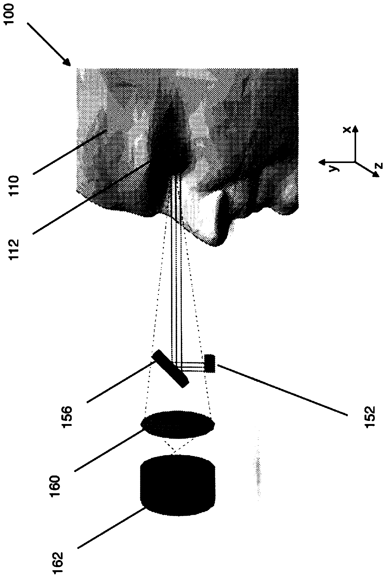
An ophthalmic device having a single or dual compartment configuration selectively emits infrared and visible light beams onto one or a pair of target eyes. The device performs eye fundus imaging andaids in the detection of ailments as indicated by anomalies in the pupillary reflex.

Description

Background technique [0001] The embodiments of the present invention generally relate to medical diagnostic devices and procedures, and more specifically to devices and procedures used in ophthalmoscopes. [0002] The medical examination of the eyes of a patient usually includes an ophthalmoscope examination, which is a method by which a medical professional (such as an ophthalmologist or a technician) examines and studies the fundus of the eye. During a typical ophthalmoscopy, medical staff will use chemical reagents to completely dilate the pupils of the patient’s eyes. Complete pupil dilation increases the field of view (FOV), which means that the opening of the fundus can be seen, imaged, and checked. The increased FOV allows medical experts to examine the internal anatomy of the eye. In the prior art, it is believed that the use of visual inspection under white light without complete pupil dilation, and the use of various devices and imaging techniques will hardly produce B...

Claims


Application Information

Patent Timeline
IPC IPC(8): A61B3/11G06V10/143G06V10/147
CPCA61B3/112G06V40/193G06V10/143G06V10/147A61B3/12A61B3/0091A61B3/0025A61B3/14G06V40/19
Inventor A.卡拉格里斯T.G.齐默尔曼
Owner IBM CORP
Learn More