
Rapid medical image processing device

A medical imaging and processing device technology, applied in the field of fast medical image processing devices, can solve problems such as fatigue of the naked eye, difficulty in observation, and easy diagnosis errors, so as to save manpower and time, avoid indistinctness and diagnostic errors with the naked eye, and reduce The effect of diagnostic error

Inactive Publication Date: 2020-07-14
王培
View PDF0 Cites 0 Cited by
  • Summary
  • Abstract
  • Description
  • Claims
  • Application Information

AI Technical Summary

Problems solved by technology

[0003] In the later stage, a video will be formed for the patient to carry and find a doctor to see the condition; generally doctors will put the video on a light board and observe the video under lighting; it not only consumes manpower, but also causes fatigue to the naked eye, and at the same time, when the light is dark, it is easy to make a wrong diagnosis Or missed diagnosis, and the smaller and thinner places are not easy to observe with the naked eye. Therefore, a fast medical image processing device is now designed

Method used


Image

  • Rapid medical image processing device
  • Rapid medical image processing device
  • Rapid medical image processing device

Examples


Embodiment

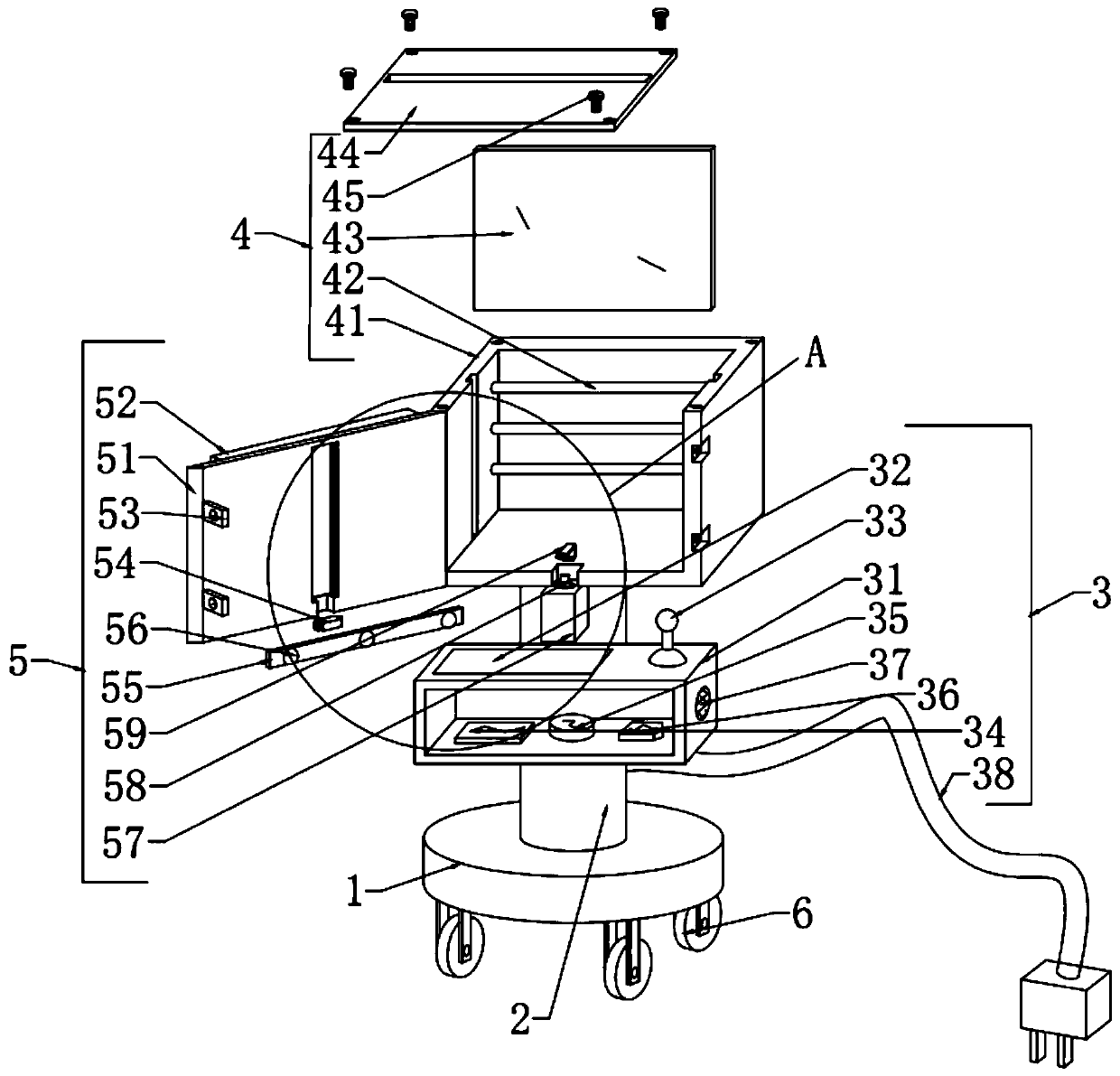
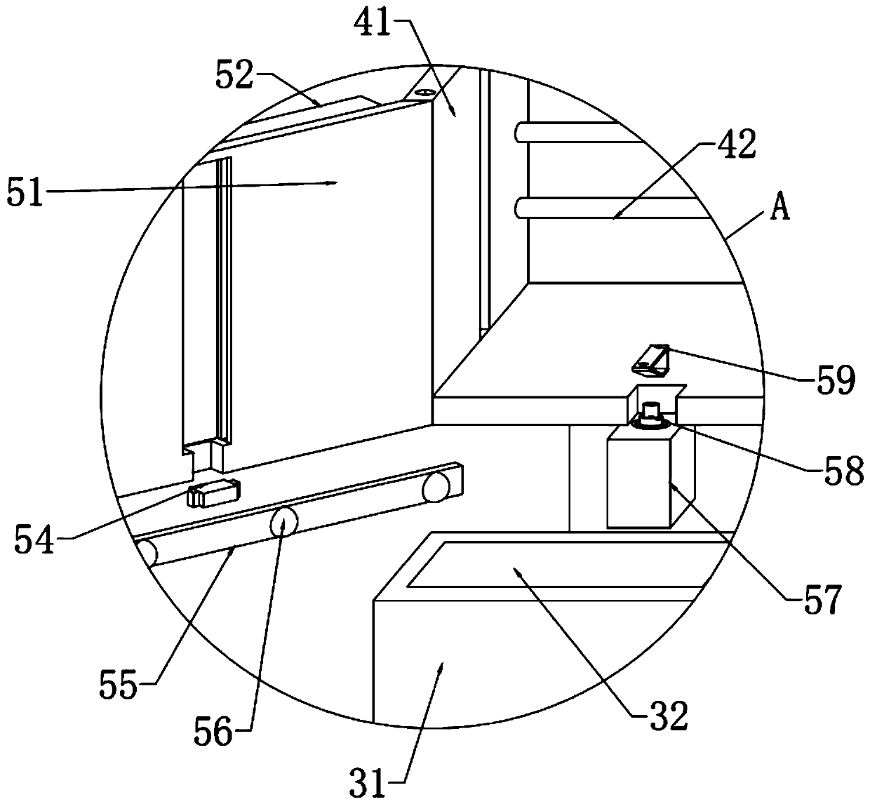
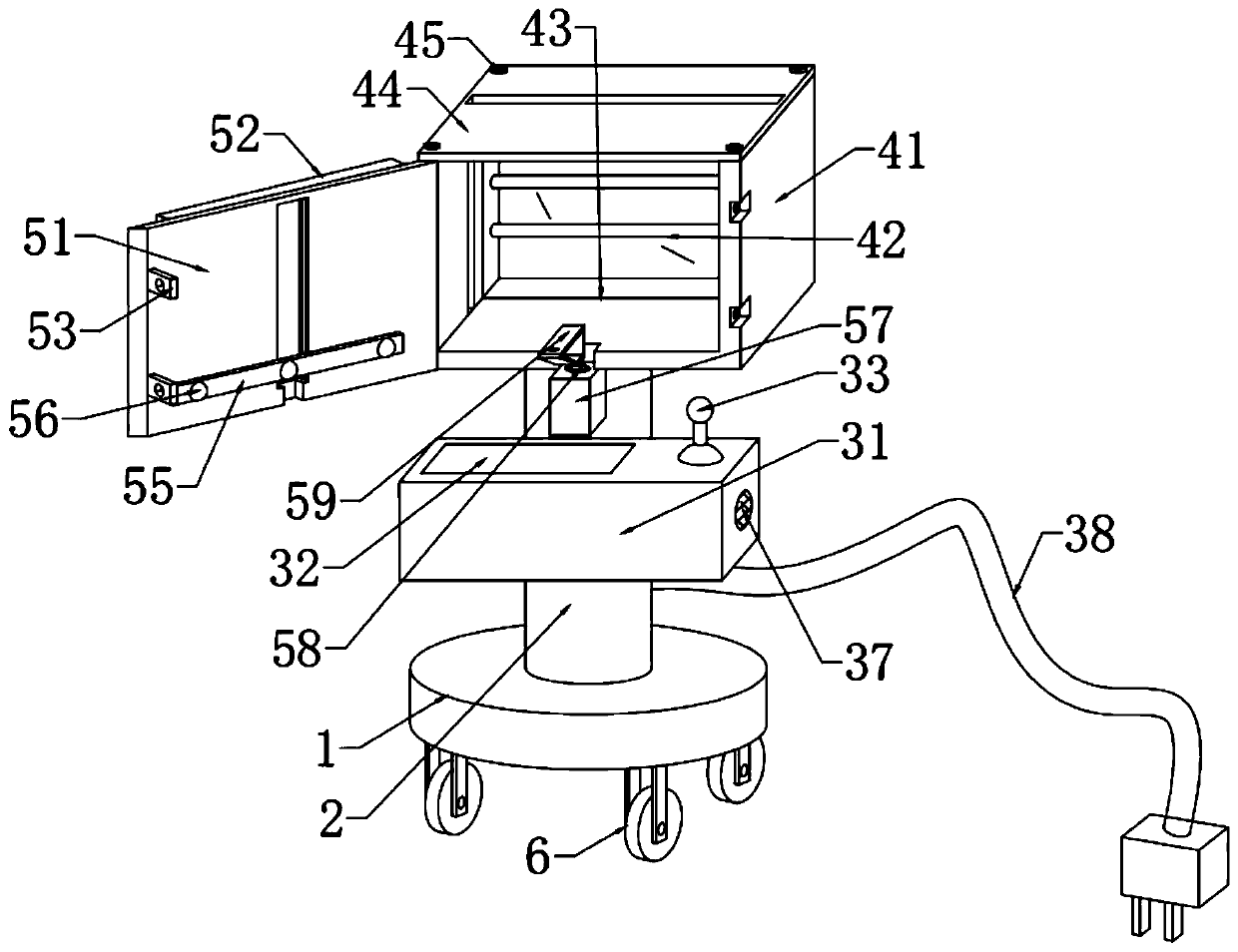
[0042] Embodiment: Attached by instruction manual Figure 1-4 It can be seen that, firstly, after the device is moved by the universal wheel 6 under the base 1, the device is energized through the power cord 38. Since the films are mostly arranged in a nine-square grid structure, one end of the CT film is inserted into the socket of the top cover 44 fixed by the bolt 45. , so that both ends are embedded in the limiting grooves on the inner side wall of the main body box 41 supported by the support rod 2, and made to fit on the baffle plate 43; , which is convenient for the equipment to scan and identify the shooting situation, and avoid medical staff's naked eyes and diagnostic errors; secondly, through the setting of the control keyboard 32 on the installation box 31 in the drive structure 3, the control panel 34 automatically controls the scanning of the carrying box in the imaging structure 5 The electric telescopic rod 58 in the 57 is driven, and the three scanning probes ...


PUM


Abstract

The invention discloses a rapid medical image processing device, which comprises a base, a support rod, a drive structure, a bearing and lighting structure and a scanning imaging structure, wherein the base is in a disc shape; one end of the support rod is fixed and welded on an upper wall of the base and the support rod is located at the central part; firstly, one end of a CT film is inserted into a socket of a head cover, and lamp tubes in the bearing and lighting structure are controlled to be illuminated for lighting, so that scanning recognition of the shooting situation by the device isfacilitated and the problems that it is difficult to distinguish the film by medical personnel through naked eyes and a diagnostic error occurs are solved; three scanning probes on a bearing frame aredriven through an electric telescopic rod to move up and down to carry out scanning distinguishing on the film and to display distinguished position and situation through a display screen, so that the manpower can be reduced, the time can be shortened, and the state of an illness and the human body shooting situation can be reflected more intuitively. The scanning probes can be controlled througha keyboard and a control rod to move up and down, and manual screening is carried out to form two-way use, so that the diagnostic error is reduced.

Description

technical field [0001] The invention relates to the field of technical medical equipment, in particular to a fast medical image processing device. Background technique [0002] Imaging technology can comprehensively and accurately obtain various quantitative and qualitative data of patients, and provide correct digital information for diagnosis, treatment planning, operation and postoperative evaluation; among them, CT is an electronic computerized tomography instrument, which uses precisely collimated X-ray Wire beams, γ-rays, ultrasonic waves, etc., together with highly sensitive detectors, perform cross-sectional scanning around a certain part of the human body one by one; this instrument has the characteristics of fast scanning time and clear images, and can be used for the examination of various diseases; [0003] In the later stage, a video will be formed for the patient to carry and find a doctor to see the condition; generally doctors will put the video on a light bo...

Claims


Application Information

Patent Timeline
Patent Type & Authority Applications(China)
IPC IPC(8): A61B6/00
CPCA61B6/462
Inventor 王培
Owner 王培
Learn More