Retinal surgery robot imaging method integrating microscope and OCT

A technology of surgical robot and imaging method, which is applied in the field of retinal surgical robot imaging, can solve the problems of poor imaging accuracy and safety of retinal surgical robot, incompatibility between accuracy and real-time performance, so as to improve the effect and success rate, and save physical exertion , the effect of reducing the risk of surgery

Active Publication Date: 2021-03-19
HARBIN INST OF TECH
View PDF6 Cites 2 Cited by
  • Summary
  • Abstract
  • Description
  • Claims
  • Application Information

AI Technical Summary

Problems solved by technology

[0005] The present invention aims to solve the problem that the existing OCT system used in surgery is not compatible with the accuracy and real-time performance due to the limitations of data processing speed and scanning mode, which leads to the problem of poor imaging accuracy and safety of retinal surgery robots.

Method used


Image

  • Retinal surgery robot imaging method integrating microscope and OCT
  • Retinal surgery robot imaging method integrating microscope and OCT
  • Retinal surgery robot imaging method integrating microscope and OCT

Examples


specific Embodiment approach 1

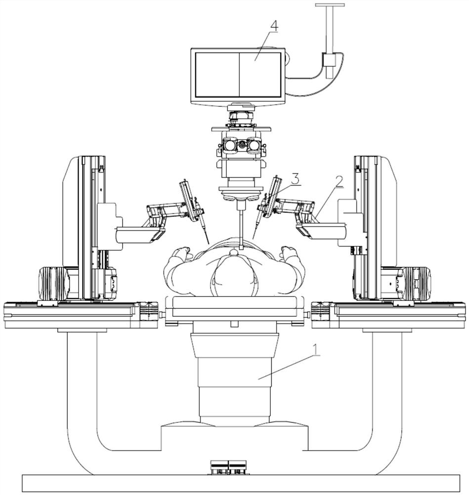
[0029] Specific implementation mode one: combine Figure 1 to Figure 4 Describe this embodiment, a kind of microscope and OCT fusion retina surgery robot imaging method, it comprises the following steps:

[0030] Step 1: Build a 3D model;

[0031] Use OCT equipment to perform C-scan on the surgical target area before the operation, and establish a three-dimensional retinal model of the treated tissue on the OCT equipment according to the three-dimensional information obtained by the C-scan, and determine the tissue structure and location information around the injection area;

[0032] Step 2: Locate the position of the needle tip 3-1;

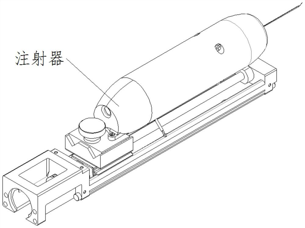
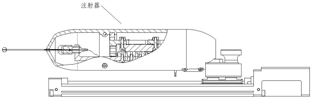
[0033] The visual information obtained by using the surgical microscope during the operation, combined with the real-time depth information fed back by the needle-integrated endoscopic OCT fiber optic probe 3-3, can be used to locate and track the position of the needle tip in real time;

[0034] Step 3: Register the 3D model of the retina in...

specific Embodiment approach 2

[0042] Specific implementation mode two: combination Figure 1 to Figure 4 Describe this embodiment. After the model is established in step 1 of this embodiment, the doctor uses the model to make a preliminary diagnosis of the lesion area, and plans the trajectory of the surgical instrument in the eye, and establishes the insertion position of the subretinal injection or vascular injection syringe. . With such arrangement, it is convenient to accurately determine the injection position. Other compositions and connections are the same as in the first embodiment.

specific Embodiment approach 3

[0043] Specific implementation mode three: combination Figure 1 to Figure 4 Describe this embodiment. During the operation in Step 2 of this embodiment, the doctor obtains the horizontal position of the syringe needle tip 3-1 relative to the target area through microscope observation, and controls the needle tip to reach above the puncture point through the main hand of the mechanical arm. At the same time, the integrated The OCT probe behind the needle tip obtains the depth information of the needle tip in real time through the A-scan function, and provides the doctor with the accurate relative distance between the needle tip and the surface of the retina. With such a setting, the contact between the needle tip and the fragile tissue in the eye is avoided, and the safety of the operation is ensured. Other compositions and connections are the same as those in Embodiment 1 or Embodiment 2.


PUM


Abstract

The invention relates to a retinal surgery robot imaging method, in particular to a retinal surgery robot imaging method integrating a microscope and OCT. According to the method, the problem that inthe prior art, a retinal surgery robot is poor in imaging accuracy and safety is solved. The method comprises the following steps of 1, performing C scanning on an operation target area by using OCT equipment before an operation, and establishing a retinal three-dimensional model of a to-be-operated tissue on the OCT equipment according to a result obtained by the C scanning; 2, positioning the position of a needle tip 3-1, and specifically combining visual information obtained by an operation microscope with real-time depth information fed back by a needle tip integrated endoscopic OCT optical fiber probe 3-3 to position and track the position of the needle tip in real time during an operation; 3, registering the retinal three-dimensional model in the step 1 and the position of the needletip in the operation; and 4, observing the penetration depth of the needle tip, and pulling out an injector after injection to finish medicine injection operation. The method is used for retinal handimaging.

Description

technical field [0001] The invention relates to a retinal surgery robot imaging method, in particular to a retinal surgery robot imaging method with microscope and OCT fusion. Background technique [0002] Eyes are delicate in structure and fragile in tissue, and most ophthalmic operations need to be performed under a microscope. Therefore, there are four problems in ophthalmic microsurgery: high risk, high difficulty, easy fatigue, and lack of subtle tactile perception. In particular, fundus and retinal surgery is not only difficult, resulting in a long learning curve for ophthalmologists, but also the precision of some surgical operations exceeds the physiological limit of human hands (the average tremor amplitude of human hands is 156 μm), making effective surgical methods impossible to achieve. Robots have the advantages of high motion precision, good stability, strong perception, and no fatigue. Therefore, in modern eye surgery, robot-assisted operations have been widel...

Claims


Application Information

Patent Timeline
Patent Type & Authority Applications(China)
IPC IPC(8): A61B34/20A61B34/37A61B34/10
CPCA61B34/20A61B34/37A61B34/10A61B2034/2065A61B2034/2055A61B2034/105
Inventor 张赫陈昱赵杰
Owner HARBIN INST OF TECH
Why Eureka
Try Eureka