
Binding elastic fixation rope system for distal tibiofibular syndesmosis separation

An elastic fixation and cable technology, applied in the field of medical devices, can solve the problems of damage to the peroneal artery and its perforating branches, and achieve the effect of solving space occupation and improving accuracy

Active Publication Date: 2021-06-15
山东大学齐鲁医院(青岛)
View PDF7 Cites 1 Cited by
  • Summary
  • Abstract
  • Description
  • Claims
  • Application Information

AI Technical Summary

Problems solved by technology

The lateral elastic fixation of the loop plate and cable system is similar to the inferior tibiofibular screw fixation. It is inserted horizontally across the tibia and fibula from about 2-3 cm above the level of the ankle joint, and the perforated branch of the peroneal artery is about 3 cm above the level of the ankle joint from back to front. Crosses the interosseous membrane, so the risk of injury to the peroneal artery and its perforating branches is greater with lateral elastic fixation

Method used


Image

  • Binding elastic fixation rope system for distal tibiofibular syndesmosis separation
  • Binding elastic fixation rope system for distal tibiofibular syndesmosis separation
  • Binding elastic fixation rope system for distal tibiofibular syndesmosis separation

Examples


Embodiment 1

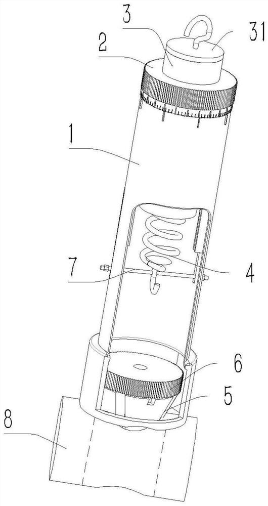
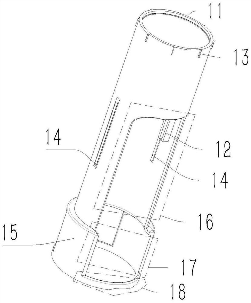
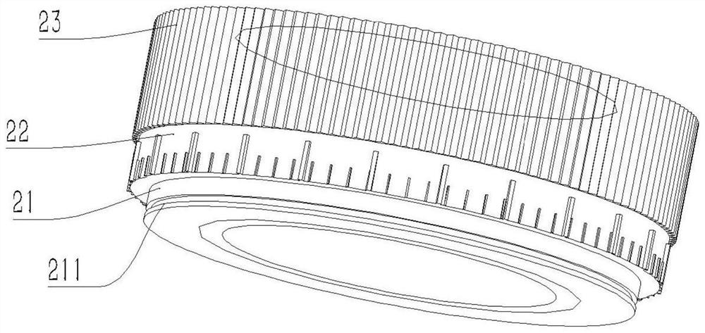
[0025] A lower tibiofibular syndesmosis separation binding elastic fixed cable system, including a cable tension measuring device and a cable fixing device, the cable tension measuring device includes a hollow cylindrical shell 1, a spring adjustment nut 2, The spring adjusts the threaded sleeve 3, the end cap 31 and the spring 4, the inner surface of one end of the housing 1 is provided with an annular protrusion 11, and the two sides of the inner surface are provided with groove-shaped guide rails 12; the side of the housing is dug 16 to facilitate hanging ropes cable; the other end of the casing 1 is inserted into the cable fastening device, and digs 17 on both sides of the cable fastening device end to facilitate screwing in the cable fastening device; The shell is placed on the surface of the bone 8 to generate an inclination; the inner side of the spring adjustment nut 2 is provided with an internal thread, and the outer surface is set in a stepped shape. The outer surfac...

Embodiment 2

[0029] On the basis of Embodiment 1, the side of the housing 1 is symmetrically provided with a strip-shaped opening 14, and one end of the spring 4 is fixed to the tightening column 7, and the two ends of the tightening column 7 pass through the strip-shaped opening 14, and set With set nut.

[0030] In order to further improve the accuracy and avoid the influence of the pulling force produced during the downward tightening of the cable on the tension of the cable, a tightening column 7 is provided. After the spring 4 adjusts the tension of the cable, the tightening column 7 fixes the spring 4, Tighten the rope at this time, effectively avoiding the influence of the pulling force on the tension.


PUM


Abstract

The invention relates to the field of medical instruments, in particular to a binding elastic fixation rope system for distal tibiofibular syndesmosis separation. The binding elastic fixation rope system comprises a rope tension measuring device and a rope fixing device. The rope tension measuring device comprises a hollow cylindrical shell, a spring adjusting nut, a spring adjusting threaded sleeve, an end cover and a spring; an annular protrusion is arranged on the inner side face of one end of the shell, and a groove-shaped guide rail is arranged on each of the two sides of the inner side face; the side face of the shell is dug to facilitate hanging of a rope. and the other end of the shell sleeves a rope fastening device, the two side faces of the shell are excavated, so that the rope fastening device can be conveniently screwed into the shell, and the shell is not broken by excavation, to prevent the shell from inclining when the shell is placed on a bone surface. According to the design of the invention, the accuracy of the tension of the binding elastic fixing rope for distal tibiofibular syndesmosis is improved, thereby solving the problem that the tension of the rope is not uniformly distributed, fixing devices at the two ends are arranged in bone cortex, thereby avoiding space occupation in soft tissue; and a more scientific basis is provided for clinical application.

Description

technical field [0001] The invention relates to the field of medical devices, in particular to a lower tibiofibular syndesmosis separation binding elastic fixation cable system. Background technique [0002] Ankle fractures associated with syndesmotic ligament injuries are relatively common clinically, and 15-23% of ankle fractures associated with syndesmotic ligament injuries require precise reduction and fixation. If the treatment is not timely or inappropriate, chronic pain, joint instability and traumatic arthritis often remain, seriously affecting the function of the ankle joint. [0003] The inferior tibiofibular screw recommended by the AO organization was first applied to the injury of the inferior tibiofibular syndesmosis. The biomechanics and clinical efficacy showed that its fixation effect was reliable, ensuring the stability of the ankle joint, but it lost the compliance of the inferior tibiofibular syndesmosis to the ankle point during exercise. Sexual adjustm...

Claims


Application Information

Patent Timeline
IPC IPC(8): A61B17/68
CPCA61B17/68
Inventor 赛佳明
Owner 山东大学齐鲁医院(青岛)
Learn More