DDAUnet-based esophageal tumor segmentation method

A tumor and esophageal technology, applied in the field of esophageal tumor segmentation based on DDAUnet, can solve the problems of esophageal tumor segmentation with few applications, limited applications, and error-prone

Pending Publication Date: 2021-07-06
山西三友和智慧信息技术股份有限公司
View PDF0 Cites 0 Cited by
  • Summary
  • Abstract
  • Description
  • Claims
  • Application Information

AI Technical Summary

Problems solved by technology

Obtaining its additional information is time-consuming, and at the same time the results are error-prone and may lead to uncertain results. In recent years, the application of deep learning in medical image analysis has attracted much attention. limited applications, and even less in esophageal tumor segmentation

Method used


Image

  • DDAUnet-based esophageal tumor segmentation method

Examples


Embodiment Construction

[0027] The technical solutions in the embodiments of the present invention will be clearly and completely described below with reference to the accompanying drawings in the embodiments of the present invention. Obviously, the described embodiments are only a part of the embodiments of the present invention, but not all of the embodiments. Based on the embodiments of the present invention, all other embodiments obtained by those of ordinary skill in the art without creative efforts shall fall within the protection scope of the present invention.

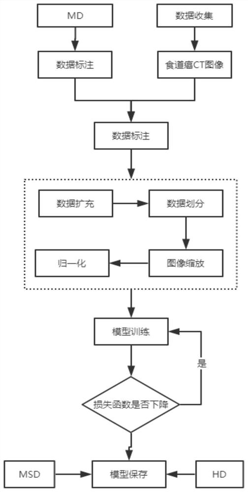
[0028] An esophageal tumor segmentation method based on DDAUnet, such as figure 1 shown, including the following steps:

[0029] S100, data collection: obtain relevant CT scan images of patients with esophageal cancer, perform image labeling, and complete the construction of data sets required for model training;

[0030] S200, data preprocessing: including data augmentation, data division, image scaling and normalization;

[0031] ...


PUM


Abstract

The invention belongs to the technical field of image segmentation, and particularly relates to a DDAUnet-based esophageal tumor segmentation method. The DDAUnet-based esophageal tumor segmentation method comprises the following steps of data acquisition, data preprocessing, model construction, model storage and model evaluation, wherein in the data acquisition, related CT scanning images of an esophageal cancer patient are acquired, image labeling is performed, and construction of a data set required by model training is completed; and the data preprocessing comprises data amplification, data division, image scaling and normalization. The invention provides a full-automatic end-to-end esophageal tumor segmentation method based on a convolutional neural network CNNs, the network is called as dilated intensive attention Unet, namely DDAUnet, space and channel attention gates in each dense block are selectively concentrated on a decisive feature map and a region, and an expanded convolutional layer is used for managing a GPU memory and increasing a network acceptance field. The method is used for image segmentation.

Description

technical field [0001] The invention belongs to the technical field of image segmentation, and in particular relates to an esophageal tumor segmentation method based on DDAUnet. Background technique [0002] Esophageal cancer is one of the least studied cancers, and it is fatal to most patients. Rapid and accurate delineation of target volume in CT images is very important for treatment and disease control. High uncertainty, especially at the cranial and caudal margins of tumors, to overcome these complexities, physicians correlate CT imaging with clinical history, endoscopic findings, endoscopic ultrasound, and other imaging modalities such as positron emission tomography Combined, however, acquiring these additional modalities is a time-consuming and expensive process. [0003] Causes of problems or deficiencies: Manual or automated delineation of esophageal tumors in CT images is very challenging. This is due to low contrast between the tumor and adjacent tissues, chang...

Claims


Application Information

Patent Timeline
Patent Type & Authority Applications(China)
IPC IPC(8): A61B6/03
CPCA61B6/032A61B6/5211
Inventor 潘晓光焦璐璐董虎弟韩丹马文芳
Owner 山西三友和智慧信息技术股份有限公司
Why Eureka
Try Eureka