
Method and device for eliminating metal artifacts of CT image, medium and electronic equipment

A metal artifact, CT image technology, applied in the field of medical imaging, can solve the problem of image resolution loss, etc., to achieve the effect of eliminating metal artifacts

Pending Publication Date: 2021-09-03
苏州晟诺医疗科技有限公司
View PDF0 Cites 0 Cited by
  • Summary
  • Abstract
  • Description
  • Claims
  • Application Information

AI Technical Summary

Problems solved by technology

[0004] However, this method of removing metal artifacts results in image loss of resolution

Method used


Image

  • Method and device for eliminating metal artifacts of CT image, medium and electronic equipment
  • Method and device for eliminating metal artifacts of CT image, medium and electronic equipment
  • Method and device for eliminating metal artifacts of CT image, medium and electronic equipment

Examples


Embodiment Construction

[0043] In order to make the purpose, technical solutions and advantages of the present disclosure clearer, the present disclosure will be further described in detail below in conjunction with the accompanying drawings. Apparently, the described embodiments are only some of the embodiments of the present disclosure, not all of them. Based on the embodiments in the present disclosure, all other embodiments obtained by persons of ordinary skill in the art without creative efforts fall within the protection scope of the present disclosure.

[0044] Terms used in the embodiments of the present disclosure are for the purpose of describing specific embodiments only, and are not intended to limit the present disclosure. The singular forms "a", "said" and "the" used in the embodiments of this disclosure and the appended claims are also intended to include plural forms, unless the context clearly indicates otherwise, "a plurality" Generally contain at least two.

[0045] It should be u...


PUM


Abstract

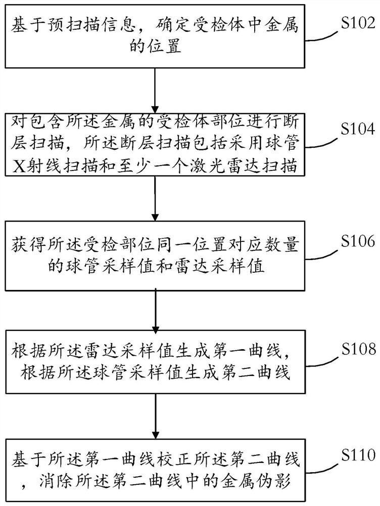
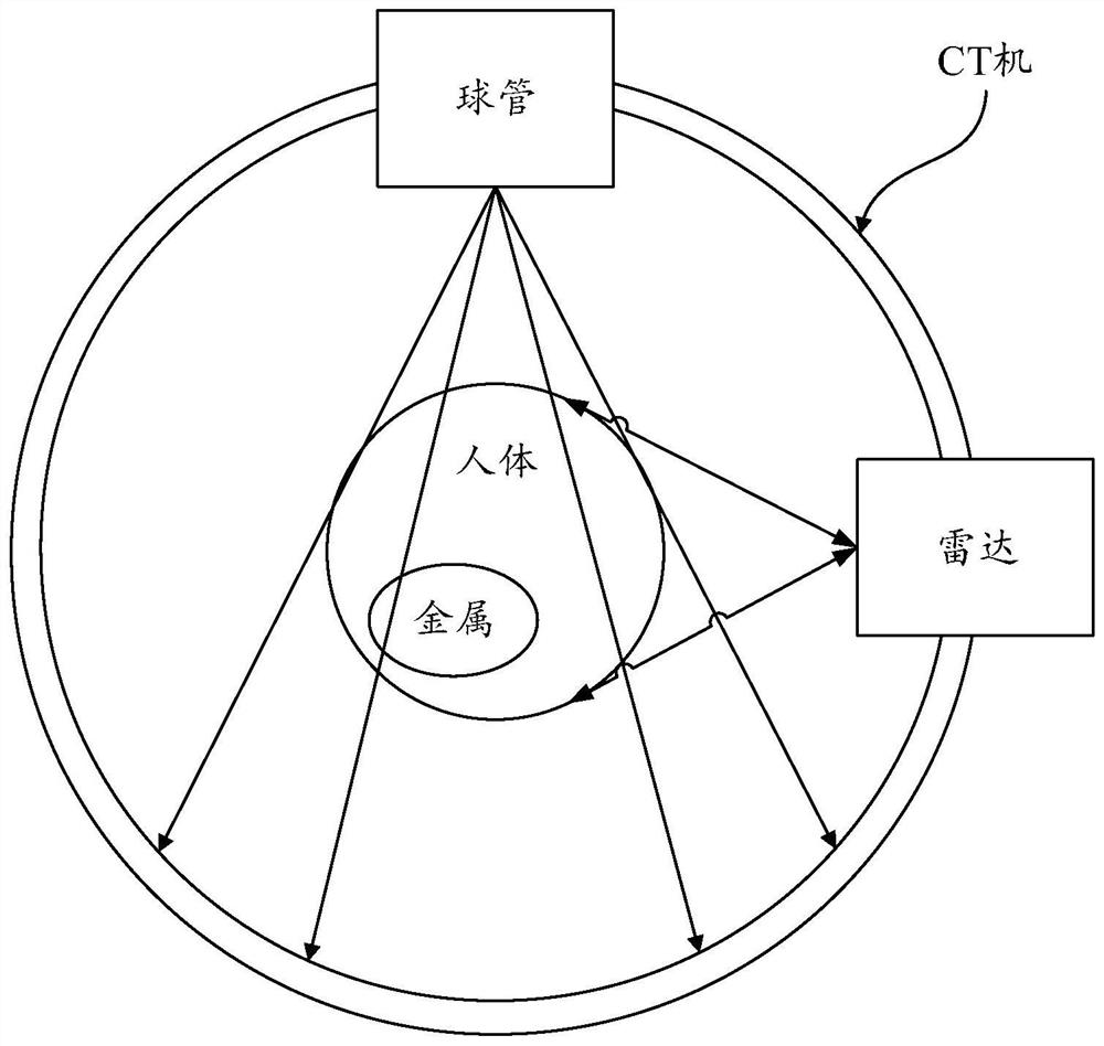
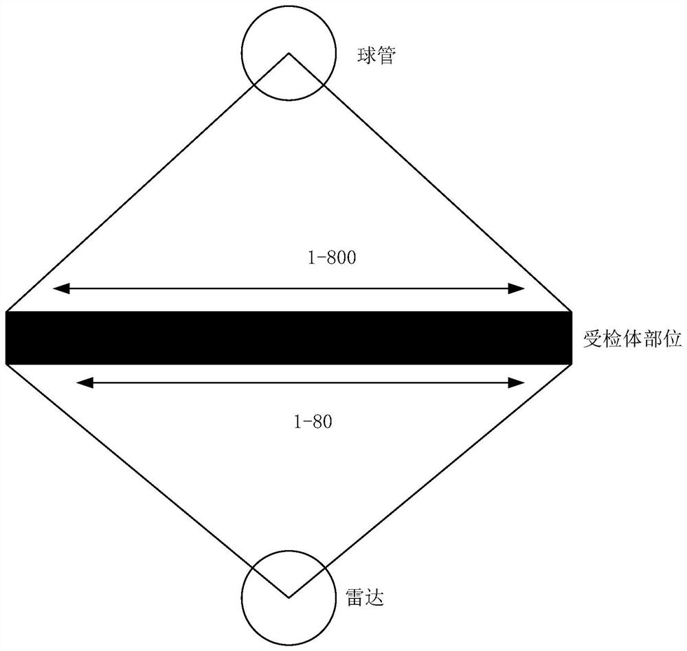
The invention provides a bulb tube sampling value correction method and device, a medium and electronic equipment. The method comprises the following steps: determining the position of metal in a subject based on pre-scanning information; carrying out tomography on the detected body part containing the metal, wherein the tomography comprises bulb tube X-ray scanning and at least one laser radar scanning; acquiring a corresponding number of bulb tube sampling values and radar sampling values at the same position of the detected part; generating a first curve according to a radar sampling value, and generating a second curve according to the bulb tube sampling value; and correcting the second curve based on the first curve, and eliminating metal artifacts in the second curve. According to the characteristics of the radar sensor, the radar sensor and the bulb tube sensor are simultaneously sampled, so the sampling positions of the radar sensor and the bulb tube sensor are completely registered; and the bulb tube sampling value including the metal artifact information is corrected by means of first curve information generated by the radar sampling value, so that the metal artifacts of the medical image generated based on the corrected bulb tube sampling value are eliminated.

Description

technical field [0001] The present disclosure relates to the field of medical imaging, in particular to a method, device, medium and electronic equipment for eliminating metal artifacts in CT images. Background technique [0002] When performing computerized tomography (Computed Tomography, CT scan for short) on a patient, if there is metal (such as a prosthesis or a stent) in the patient's body, there will be metal artifacts in the CT image, which will affect the doctor's diagnosis. [0003] At present, for the correction algorithm of metal artifacts in CT images, usually, according to the CT value obtained in the CT image, the metal area is segmented, and the data is corrected by front and back projection, and then the calibrated data replaces the data in the metal area . [0004] However, this method of removing metal artifacts results in a loss of image resolution. Therefore, the present disclosure proposes a method for correcting the sampling value of the bulb to solv...

Claims


Application Information

Patent Timeline
Patent Type & Authority Applications(China)
IPC IPC(8): G06T11/00G06T7/33
CPCG06T11/008G06T7/33G06T2207/10081G06T2207/10044
Inventor 任毅
Owner 苏州晟诺医疗科技有限公司
Learn More