Coronary artery segmentation method and device based on multi-slice combination

A coronary artery and multi-slice technology, applied in the field of coronary artery segmentation based on multi-slice combination, can solve problems such as inaccurate segmentation results of three-dimensional cardiovascular images, and achieve the effect of improving segmentation accuracy and accuracy

Active Publication Date: 2021-09-17
GUANGDONG UNIV OF TECH
View PDF9 Cites 2 Cited by
  • Summary
  • Abstract
  • Description
  • Claims
  • Application Information

AI Technical Summary

Problems solved by technology

[0004] In order to overcome the defect of inaccurate segmentation results of three-dimensional cardiovascular images in the prior art, the present invention provides a coronary artery segmentation method and device based on multi-slice combination, Neighborhood information is fully considered to improve the accuracy of segmentation results

Method used


Image

  • Coronary artery segmentation method and device based on multi-slice combination
  • Coronary artery segmentation method and device based on multi-slice combination
  • Coronary artery segmentation method and device based on multi-slice combination

Examples


Embodiment 1

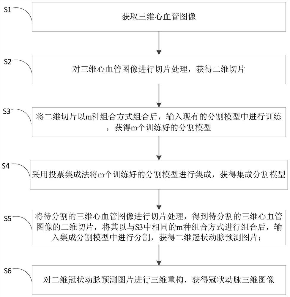
[0050] This embodiment provides a coronary artery segmentation method based on multi-slice combination, such as figure 1 As shown, the method includes the following steps:

[0051] S1: acquiring a three-dimensional cardiovascular image;

[0052] The three-dimensional cardiovascular images are obtained by CT or MRI techniques. Both CT and MRI techniques are non-invasive imaging techniques with high safety.

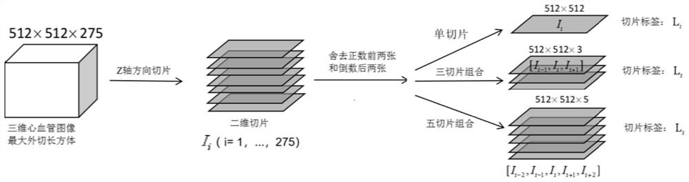
[0053] S2: Perform slice processing on the three-dimensional cardiovascular image to obtain two-dimensional slices;

[0054] Construct the largest circumscribed cuboid for the three-dimensional cardiovascular image, the size of which is a*b*c, where a, b, and c are the length, width, and height values ​​of the largest circumscribed cuboid of the three-dimensional cardiovascular image; the maximum circumscribed cuboid of the three-dimensional cardiovascular image The cuboid is placed in the three-dimensional coordinate system, sliced ​​​​in the Z-axis direction, and a two...

Embodiment 2

[0087] This embodiment provides a coronary artery segmentation device based on multi-slice combination, such as Figure 4 As shown, the device includes:

[0088] An image acquisition module, configured to acquire a three-dimensional cardiovascular image;

[0089] A slicing module, configured to perform slicing processing on a three-dimensional cardiovascular image to obtain a two-dimensional slice;

[0090] The combined training module is used to combine the two-dimensional slices with m kinds of combinations, and then input them into the segmentation model for training to obtain m trained segmentation models;

[0091] A model integration module is used to integrate m trained segmentation models to obtain an integrated segmentation model;

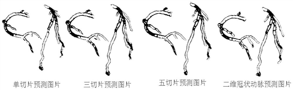
[0092] The image input module to be segmented is used to perform slice processing on the three-dimensional cardiovascular image to be segmented to obtain a two-dimensional slice of the three-dimensional cardiovascular image to be segmente...


PUM


Abstract

The invention discloses a coronary artery segmentation method and device based on multi-slice combination. The method comprises the following steps: acquiring a three-dimensional cardiovascular image; slicing the three-dimensional cardiovascular image to obtain a two-dimensional slice; combining the two-dimensional slices in m combination modes, and inputting the combined two-dimensional slices into segmentation models for training to obtain m trained segmentation models; integrating the m trained segmentation models to obtain an integrated segmentation model; combining the two-dimensional slices of the three-dimensional cardiovascular image to be segmented in the same m combination modes, and inputting the combined two-dimensional slices into the integrated segmentation model for segmentation to obtain a two-dimensional coronary artery prediction picture; and performing three-dimensional reconstruction on the two-dimensional coronary artery prediction picture to obtain a coronary artery three-dimensional image. According to the method, the neighborhood information between the two-dimensional slices is fully considered, the deficiency of the neighborhood information during input in a single slice mode is made up, cross-slice consistency prediction is carried out, and the accuracy of a coronary artery segmentation result is improved.

Description

technical field [0001] The present invention relates to the technical field of coronary artery segmentation, more specifically, to a method and device for coronary artery segmentation based on multi-slice combination. Background technique [0002] Coronary artery disease is one of the most common cardiovascular diseases, and the segmentation of coronary arteries is of great significance for the accurate location and quantitative analysis of cardiovascular diseases. In recent years, the development of non-invasive imaging technology has revolutionized the accuracy of diagnosis, and modern equipment can image internal organs in submillimeter detail. With the development of computer technology, three-dimensional imaging has been realized, and it is widely used in computed tomography of the heart to identify internal organs according to their intensity. In computed tomography of the heart, congested vessels appear brighter than surrounding tissue, allowing manual tracking of co...

Claims


Application Information

Patent Timeline
Patent Type & Authority Applications(China)
IPC IPC(8): G06T17/00G06T7/11G06T5/50G06T7/90G06N3/04G06N3/08
CPCG06T17/00G06T7/11G06T5/50G06T7/90G06N3/04G06N3/08G06T2207/10081G06T2207/10088G06T2207/20021G06T2207/20081G06T2207/20221G06T2207/30101
Inventor 曾安米晨晰甘孟坤吴春彪潘丹
Owner GUANGDONG UNIV OF TECH
Why Eureka
Try Eureka