An ultrasound image segmentation method for thyroid nodules based on cascaded total convolution neural network

A convolutional neural network, thyroid nodule technology, applied in neural learning methods, biological neural network models, image analysis, etc. The effect of high segmentation accuracy

Active Publication Date: 2018-12-25
TIANJIN UNIV
View PDF2 Cites 38 Cited by
  • Summary
  • Abstract
  • Description
  • Claims
  • Application Information

AI Technical Summary

Problems solved by technology

However, due to the characteristics of low contrast, speckle echo, blurred border of thyroid nodules, and calcification point shadows

Method used


Image

  • An ultrasound image segmentation method for thyroid nodules based on cascaded total convolution neural network
  • An ultrasound image segmentation method for thyroid nodules based on cascaded total convolution neural network
  • An ultrasound image segmentation method for thyroid nodules based on cascaded total convolution neural network

Examples


Embodiment 1

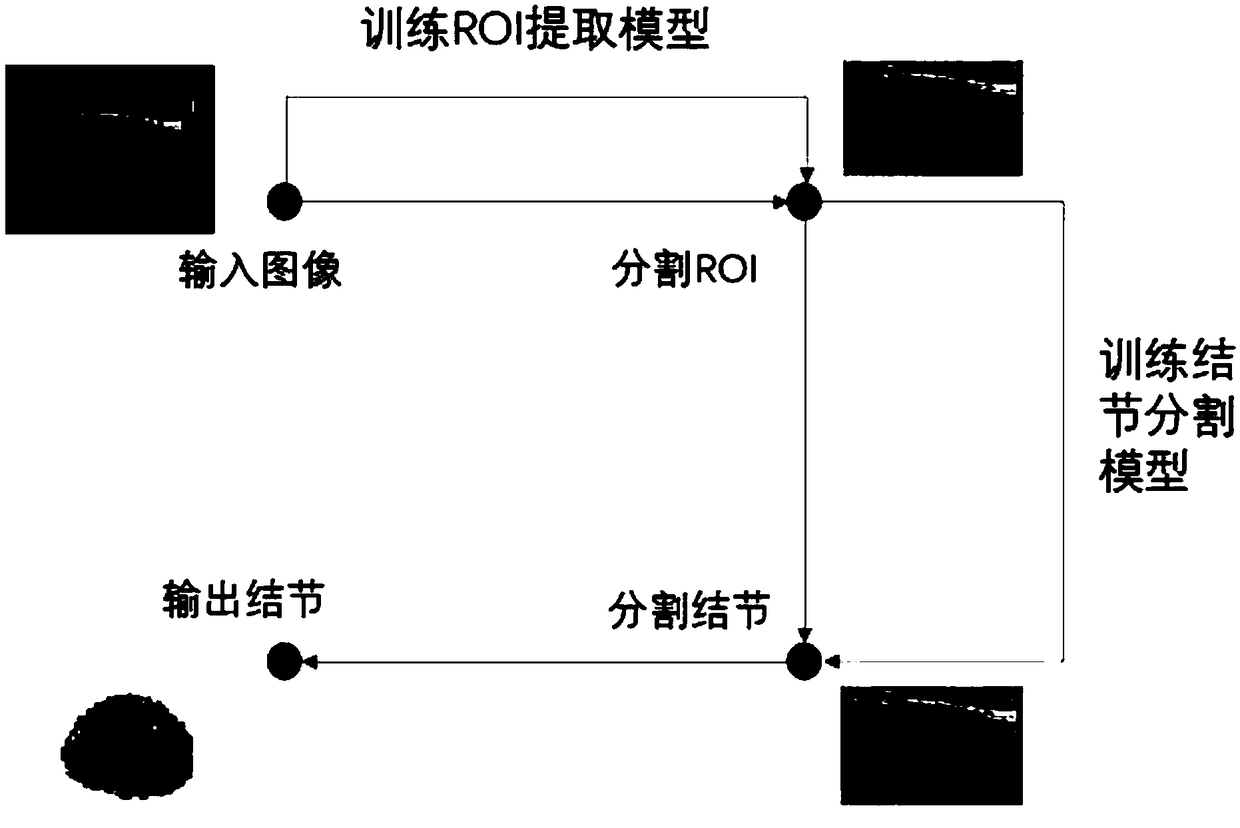
[0025] A cascaded fully convolutional neural network segmentation method for ultrasound images of thyroid nodules, see figure 1 , the method includes the following steps:

[0026] 101: Construct a simple fully convolutional neural network based on U-Net, segment the ultrasound image in the thyroid ultrasound data according to the simple fully convolutional neural network, and segment the region of interest from it;

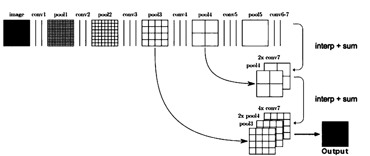
[0027] 102: Use the VGG19-FCN network as the downsampling layer to extract the deep features of the region of interest, so as to realize the automatic semantic segmentation of thyroid nodules;

[0028] Wherein, the simple and easy fully convolutional neural network in step 102 includes:

[0029] Five convolutional layers for downsampling, and five upsampling layers for upsampling;

[0030] Among them, the first five convolution conv are composed of two 3x3 convolution layers and one pooling layer, each convolution layer uses ReLU as the activation function, and ...

Embodiment 2

[0037] In order to achieve the above purpose, the technical idea of ​​the embodiment of the present invention is: read the thyroid ultrasound data, remove the background area in the ultrasound image through a simple U-shaped fully convolutional neural network, construct training and test sample data sets, and construct a deep full volume The product neural network, namely FCN, is trained on the data set to obtain the semantic segmentation results suitable for ultrasound images. The specific steps include:

[0038] 201: Read the thyroid ultrasound data, and remove the background area of ​​the ultrasound image through a simple full convolutional neural network based on U-Net:

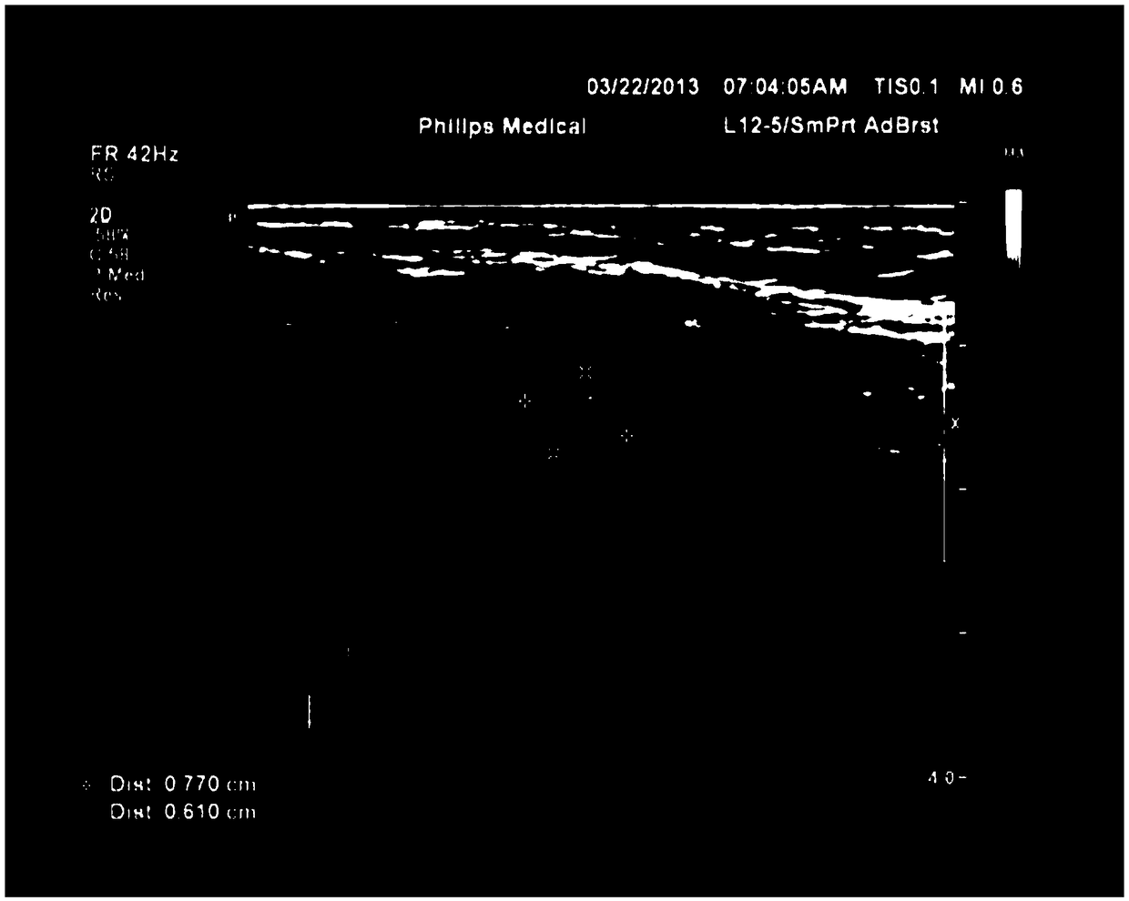
[0039] Among them, the ultrasound image is composed of a region of interest (ROI) and a background area. The ROI contains important diagnostic information, and the background area contains a large number of highlighted letters and symbols, which easily cause interference when the neural network extracts th...

Embodiment 3

[0067] Combine below Figure 1-Figure 3 The scheme in embodiment 1 and 2 is further introduced, see the following description for details:

[0068] 1) Read the ultrasound image data of thyroid nodules, which can be in various types of image formats:

[0069] 2) First read all the pictures in the training set, including ultrasound images and ROI-marked label data images, and train a UNET-based automatic ROI segmentation model (ie, the first part of the neural network);

[0070] 3) Using the segmented ROI and labeled data images of thyroid nodules under the guidance of experts as input, a model for automatic segmentation of thyroid nodules based on VGG19-FCN is trained, and the ROI segmentation model and nodule segmentation model are cascaded to form automatic segmentation The medical aid device for ultrasound images of thyroid nodules is then read into the test set data for testing. When using this method to automatically segment thyroid nodules, it is only necessary to read ...


PUM


Abstract

The invention discloses an ultrasonic image segmentation method of cascaded total convolution neural network for thyroid nodule, which comprises the following steps of: constructing an ultrasonic image segmentation method based on U-NET, the region of interest (ROI) is segmented from the ultrasound images of thyroid gland based on the simplified total convolution neural network (TCNN); using the VGG19-FCN network as the down-sampling layer to extract the deep features of ROI, so as to realize the automatic semantic segmentation of thyroid nodules. The simplified total convolution neural network comprises a fifth convolution layer for downsampling and a fifth upsampling layer for upsampling. The first five convolutions conv are composed of two 3x3 convolution layers and one pooling layer, each convolution layer uses ReLU as the activation function, and the last five upsampling layers are deconvolution layers. The invention provides a high-precision nodule image for identifying benign and malignant thyroid nodules, thereby playing a better auxiliary role in medical diagnosis.

Description

technical field [0001] The present invention relates to the field of image processing technology and the field of medical aided diagnosis, in particular to an image semantic segmentation method, in particular to a method for automatically segmenting nodules in thyroid ultrasound images based on fully convolutional neural networks. Background technique [0002] Thyroid nodules are a common disease now. According to surveys, the incidence of thyroid nodules in the population is nearly 50%, but only 4%-8% of thyroid nodules can be palpated in clinical palpation. Thyroid nodules can be divided into benign and malignant, and the incidence of malignancy is 5%-10%. Early detection of lesions is of great significance for distinguishing benign from malignant, clinical treatment and surgical selection. Ultrasound examination of thyroid nodules based on ultrasound imaging technology is a common way of examination at present. However, the results of doctors' diagnostic ultrasound thyro...

Claims


Application Information

Patent Timeline
IPC IPC(8): G06T7/181G06T7/11G06N3/08G06N3/04
CPCG06N3/08G06T7/11G06T7/181G06N3/045
Inventor 应翔尉智辉于健赵满坤徐天一高洁
Owner TIANJIN UNIV
Why Eureka
Try Eureka