
Automatic segmentation method for rectal cancer CT image based on U-Transformer

A CT image and automatic segmentation technology, applied in image analysis, neural learning methods, image data processing, etc., can solve problems such as instability and low efficiency of manual segmentation, achieve high-precision medical segmentation, realize medical segmentation, and avoid low efficiency. and unstable effects

Active Publication Date: 2021-11-19
ZHEJIANG UNIV OF FINANCE & ECONOMICS
View PDF9 Cites 9 Cited by
  • Summary
  • Abstract
  • Description
  • Claims
  • Application Information

AI Technical Summary

Problems solved by technology

[0005] In order to overcome the deficiencies in the prior art, the present invention aims to propose a method for automatic segmentation of rectal cancer CT images based on U-Transformer, which realizes the segmentati

Method used


Image

  • Automatic segmentation method for rectal cancer CT image based on U-Transformer
  • Automatic segmentation method for rectal cancer CT image based on U-Transformer
  • Automatic segmentation method for rectal cancer CT image based on U-Transformer

Examples


Embodiment 1

[0085] Embodiment 1: Data preprocessing module: preprocessing to experimental data

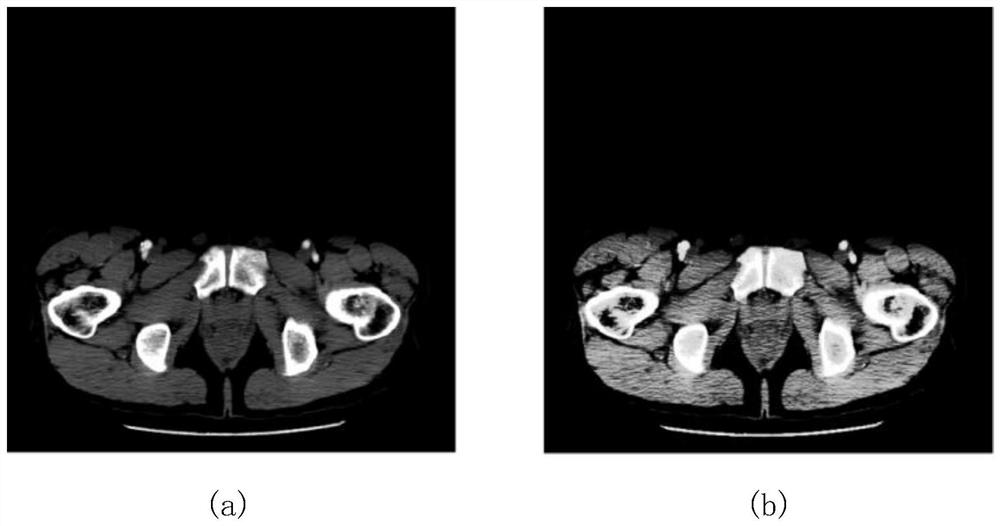
[0086] (1) Perform histogram equalization and normalization processing on the data, such as figure 1 shown.

[0087] (2) Carry out data regulation on CT images and build a protocol database.

[0088] (3) Carry out geometric transformation methods such as rotation, mirror image, and horizontal flip on CT images, and perform data enhancement and amplify training samples to reduce overfitting.

[0089] (4) Unify the size of each CT image and marker map.

[0090] (5) Divide the data set into training set, verification set and test set according to the ratio of 0.8:0.1:0.1.

Embodiment 2

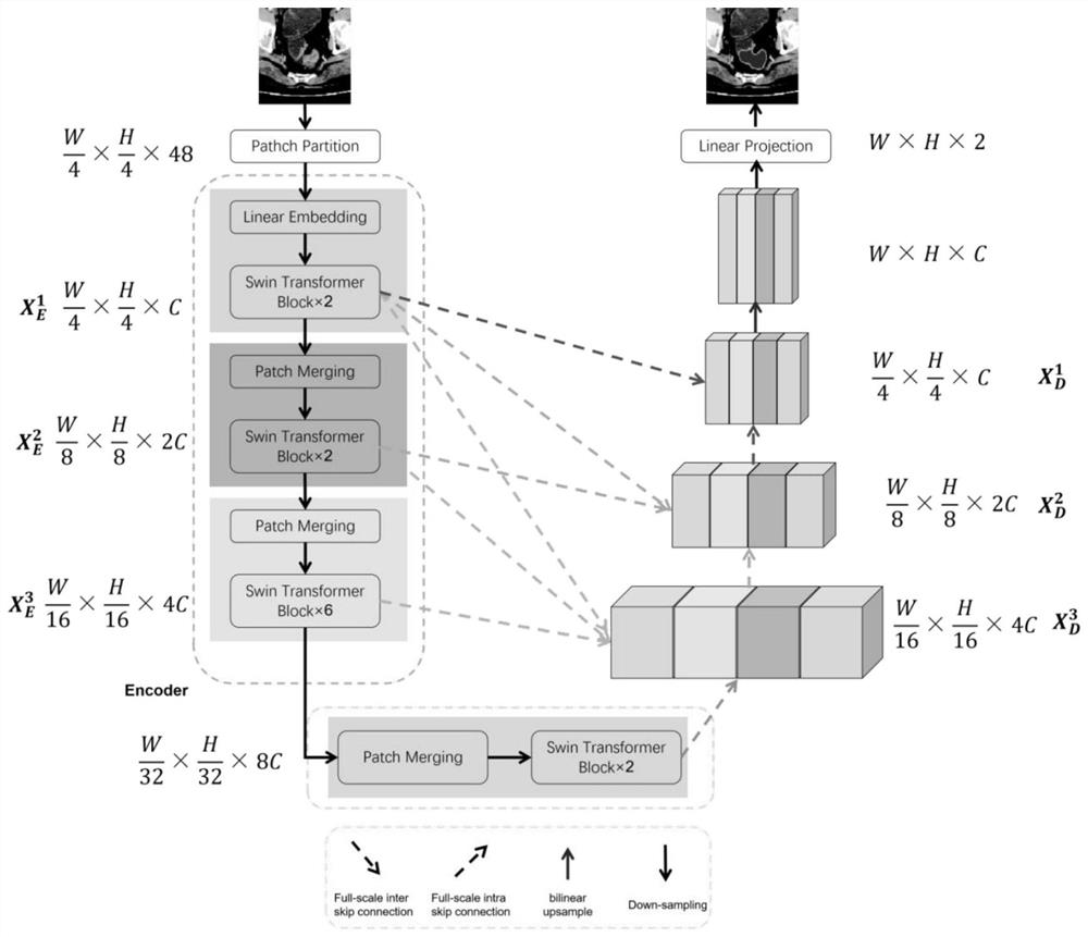
[0091] Embodiment 2: U-Transformer network model construction module: construct U-Transformer network model.

[0092] (1) Construct the Patch Embedding layer. Transform the two-dimensional CT image to obtain M pieces of size P 2 One-dimensional patch embeddings of C.

[0093] (2) Build the Swin Transformer Block. The specific Swin Transformer Block such as Figure 4 Shown: 1. Use the W-MSA mechanism to calculate the self-attention score inside the window 2. Use the SW-MSA mechanism to calculate the attention score between different windows.

[0094] (3) Build Patch Merging. The specific Patch Merging performs interval sampling on the H and W dimensions and stitches them together to achieve the purpose of downsampling.

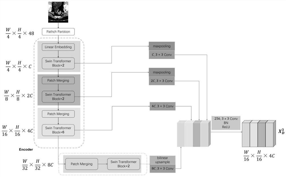
[0095] (4) Construct full-scale skip connections. The specific full-scale skip connection mechanism is as follows: image 3 Shown: 1. For high-level semantic features, we first use maximum pooling to reduce the size of its feature map, and then use a 3×3...

Embodiment 3

[0098] Embodiment 3: U-Transformer network model training module: training U-Transformer network model

[0099] (1) The Adam optimization method is used as the optimization method, and the cross entropy is used as the loss function for training. The cross-entropy formula is as follows:

[0100]

[0101] Among them, y i is the value of the pixel point i in the real eye mask diagram of the table note, and the value is 0 or 1; is the value of pixel i in the mask image obtained by the algorithm, and the value range is 0 to 1; N is the total number of pixels in the segmentation image and the annotation mask image.

[0102] (2) Pre-train the U-Transformer network model on the CIFAR-100 dataset.


PUM


Abstract

The invention discloses an automatic segmentation method for a rectal cancer CT image based on a U-Transformer. The method comprises the following steps: 1, preprocessing experimental data; 2, constructing a U-Transformer network model; 3, training the U-Transformer network model; 4, adopting the trained U-Transformer network model to carry out segmentation of the rectal cancer in the CT image, and evaluating the segmentation effect. According to the invention, the segmentation of the CT image of the rectal cancer tumor is realized, and the defects of low efficiency and instability of manual segmentation are avoided, so that an accurate basis is provided for diagnosis, treatment and operation guidance of related diseases. Compared with other U-shaped network structures, the method can learn global features and has a larger visual perception range, so that high-precision medical segmentation is realized.

Description

technical field [0001] The invention relates to a method for automatic segmentation of CT images of rectal cancer tumors based on U-Transformer, belonging to the technical field of precise segmentation of rectal cancer. Background technique [0002] In 2018, the incidence and mortality of rectal cancer ranked fourth among all cancers worldwide. According to clinical medical guidelines, the survival and prognosis of rectal cancer patients are highly correlated with tumor stage. But in general, most symptomatic patients develop to advanced stage, and the 5-year survival rate of advanced stage patients is much lower than that of early stage patients. Early detection of tumors is very important to improve the survival time of patients. [0003] At present, the early screening methods for rectal cancer mainly include: fecal occult blood test, colonoscopy and medical imaging examination. Occult blood substances are easy to obtain in the detection and identification, but the ide...

Claims


Application Information

Patent Timeline
IPC IPC(8): G06T7/00G06T7/11G06N3/04G06N3/08
CPCG06T7/0012G06T7/11G06N3/08G06T2207/10081G06T2207/20081G06T2207/20084G06T2207/30096G06T2207/30028G06N3/045Y02A90/10
Inventor 宋海裕王浩宇吴海燕张志强邓胜春冯小青陈琰宏彭娟娟
Owner ZHEJIANG UNIV OF FINANCE & ECONOMICS
Learn More