Kidney image segmentation method based on shape prior

An image segmentation and kidney technology, applied in the field of image processing, can solve the problems of uneven grayscale morphology of kidney tissue, affecting the segmentation accuracy of the level set method, and achieve the effect of solving automatic segmentation problems and improving segmentation accuracy.

Inactive Publication Date: 2021-11-30
CARBON (SHENZHEN) MEDICAL DEVICE CO LTD
View PDF0 Cites 2 Cited by
  • Summary
  • Abstract
  • Description
  • Claims
  • Application Information

AI Technical Summary

Problems solved by technology

However, the gray scale of kidney tissue is not uniform, the gray scale of each organ is close, the adjacent organs are connected, and they are susceptible to morphological changes of lesions, which will affect the segmentation accuracy of the level set method.

Method used


Image

  • Kidney image segmentation method based on shape prior
  • Kidney image segmentation method based on shape prior
  • Kidney image segmentation method based on shape prior

Examples


Embodiment Construction

[0042] The following will clearly and completely describe the technical solutions in the embodiments of the present invention with reference to the accompanying drawings in the embodiments of the present invention. Obviously, the described embodiments are only some, not all, embodiments of the present invention. Based on the embodiments of the present invention, all other embodiments obtained by persons of ordinary skill in the art without creative work, any modifications, equivalent replacements, improvements, etc., shall be included in the protection scope of the present invention Inside.

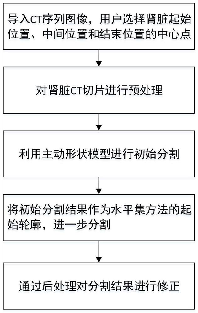
[0043] Such as Figure 1 to Figure 3 As shown, this embodiment discloses a kidney image segmentation method based on shape prior, including the following steps:

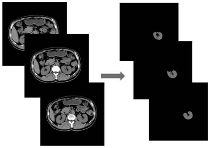
[0044] S1. Import a human body CT sequence image including the kidney, and the user selects the kidney center point of the start position, middle position and end position of the kidney slice;

[0045] S2. Preprocessing the ki...


PUM


Abstract

The invention discloses a kidney image segmentation method based on shape prior, and the method comprises the following steps: S1, importing a human CT sequence image containing a kidney, and selecting kidney center points of a starting position, a middle position and an ending position of a kidney slice by a user; S2, performing pretreatment on the kidney CT slice, wherein the pretreatment comprises bed removal, denoising and enhancement; S3, based on a result of the step S1, performing template training by using an active shape model, finally performing initial segmentation, and taking a segmentation result as an initial segmentation contour; S4, based on a result of the step S3, taking the initial segmentation contour as an initial contour of a level set method, and carrying out further segmentation; and S5, based on the segmentation result in the step S4, performing post-processing by using the front and back continuous attributes of the CT sequence image, and correcting the segmentation result. The shape template is generated through training of a small number of kidney CT sequence images, so that the kidney image is reasonably segmented, and the segmentation precision is improved.

Description

technical field [0001] The invention belongs to the technical field of image processing, in particular to a kidney image segmentation method based on shape prior. Background technique [0002] Kidney CT sequence image segmentation is an important prerequisite for quantitative analysis of kidney diseases, and it plays a supporting role in the diagnosis and treatment of diseases. It is also an important means of three-dimensional visualization of organ and tissue images in computer-aided diagnosis, virtual surgery, and biological system simulation. Since the gray scale of each organ in the abdominal CT image is similar, the adjacent organs are connected and affected by the partial volume effect. On the other hand, the tissue structure of the kidney is complex and has various shapes, and related diseases will also lead to significant changes in the shape of the kidney. Therefore, automatic segmentation of kidney images has become a difficult point in medical image segmentation....

Claims


Application Information

Patent Timeline
Patent Type & Authority Applications(China)
IPC IPC(8): G06T7/66G06T7/62G06T7/187G06T7/13G06T7/11G06T5/00G06K9/62
CPCG06T7/66G06T5/002G06T7/11G06T7/13G06T7/187G06T7/62G06T2207/10081G06T2207/30084G06F18/214
Inventor 吴梦麟章世平
Owner CARBON (SHENZHEN) MEDICAL DEVICE CO LTD
Why Eureka
Try Eureka