Image division method aiming at dynamically intensified mammary gland magnetic resonance image sequence

A technology of dynamic enhancement and magnetic resonance, which is applied in image analysis, image data processing, medical science, etc., can solve the problems of long segmentation operation time, increased workload, interference image comparison analysis, etc., to improve stability and reduce complexity The effect of speeding up the performance and splitting speed

Inactive Publication Date: 2008-12-31
TSINGHUA UNIV
View PDF0 Cites 63 Cited by
  • Summary
  • Abstract
  • Description
  • Claims
  • Application Information

AI Technical Summary

Problems solved by technology

[0009] To sum up, there are several difficulties in the segmentation of dynamically enhanced breast magnetic resonance images: First, the segmentation of 3D data at a single time point with blurred contours is difficult to automatically realize, and often requires 3D display and human-computer interaction. Increased workload; second, due to the large amount of image data, the segmentation calculation time is very long; third, the boundary between the thorax and breast is not clear in the image, and the pulsation of internal organs in the thorax seriously interferes with different time points. Therefore, it is very difficult and very important to accurately separate the breast and chest cavity

Method used


Image

  • Image division method aiming at dynamically intensified mammary gland magnetic resonance image sequence
  • Image division method aiming at dynamically intensified mammary gland magnetic resonance image sequence
  • Image division method aiming at dynamically intensified mammary gland magnetic resonance image sequence

Examples


Embodiment Construction

[0060] The method of the present invention will be described in further detail below in conjunction with the accompanying drawings and examples.

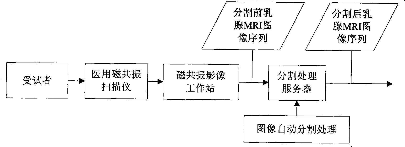
[0061] Such as figure 1 As shown, the input image sequence of the present invention is obtained from the image workstation of the medical magnetic resonance scanner, and the image workstation transmits the dynamically enhanced breast magnetic resonance image of the examinee to the image segmentation system of the present invention for operation. On the computer, the image segmentation method and system for the dynamic enhanced breast magnetic resonance image sequence of the present invention are processed to obtain the segmentation result of the sequence image.

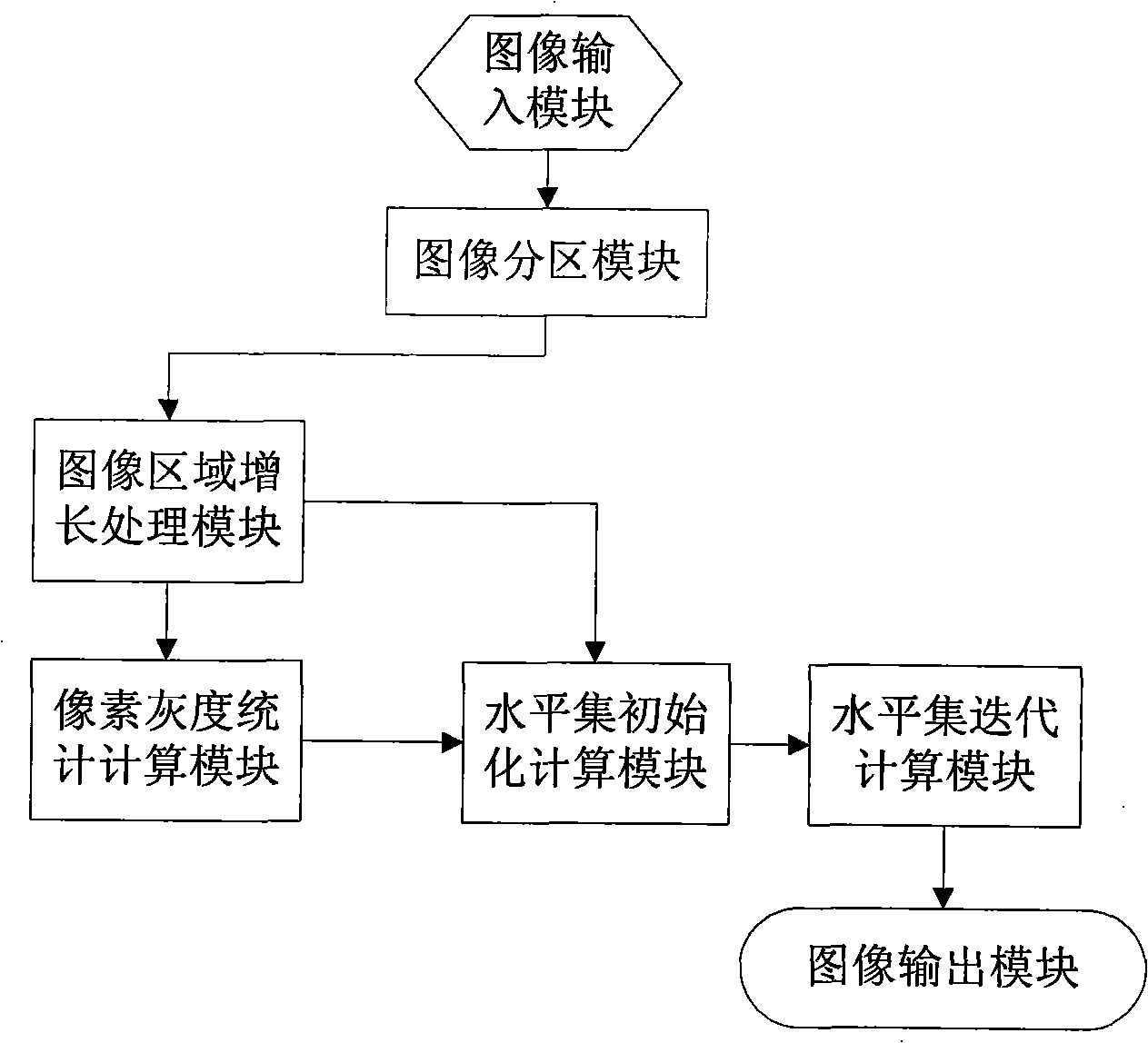
[0062] In order to implement the processing method of the present invention, it is necessary to construct such as figure 2The processing modules shown include an image input module, an image partition module, an image region growth processing module, a pixel gray level st...


PUM


Abstract

The invention discloses an image segmentation method for a dynamic contrast-enhanced mammary gland MRI sequence, pertaining to the field of magnetic resonance image processing techniques, which is characterized by comprising the following steps: a three-dimensional magnetic resonance image sequence of the section of the mammary gland is put into a computer; the image is divided into two parts including a mammary gland-air interface and a mammary gland-chest interface; a breast-air boundary is obtained by a splitting transaction in which a dynamic threshold controls the regional growth; an initial profile of the mammary gland and the chest is obtained in the same way, the complex profile of the breast and the chest is obtained with a method of controlling a level set; a three-dimensional magnetic resonance image sequence of a point-in-time is obtained by split jointing the segmentation results and taken as an initial position of the next group three-dimensional image segmentation. The image segmentation method of the invention increases the segmentation speed, solves the problem that a level set algorithm can not easily determine the initial profile and the velocity function and realizes an automatic segmentation of the complex dynamic contrast-enhanced mammary-gland magnetic resonance image with plenty of data.

Description

technical field [0001] The solution of the invention belongs to the application field of computer-aided analysis of medical images, and in particular relates to a method for segmenting dynamically enhanced breast magnetic resonance image sequences. Background technique [0002] Breast cancer is one of the most common malignant tumors in middle-aged and elderly women. It has already ranked first in female malignant tumors in China, and has a tendency of increasing year by year and younger incidence. At present, there is no good strategy for the prevention of breast cancer, so early diagnosis and determination of tumor nature are the most effective ways to improve the cure rate and reduce the mortality rate. The detection and analysis of breast images is an important basis for the successful detection, diagnosis, and treatment of breast tumors. In the past two decades, mammography has been widely used in clinical practice to detect early asymptomatic breast tumors. However, du...

Claims


Application Information

Patent Timeline
Patent Type & Authority Applications(China)
IPC IPC(8): G06T7/00A61B5/055
Inventor 王广志丁辉王琪
Owner TSINGHUA UNIV
Why Eureka
Try Eureka