Focus segmentation method and device and medium

A lesion and data set technology, applied in the field of computer vision, can solve problems such as model mis-segmentation, restriction screening diagnosis, mis-segmentation, etc., and achieve the effect of overcoming the problems of mis-segmentation, mis-segmentation, and unbalanced scene pixels

Pending Publication Date: 2022-04-08
北京理工大学重庆创新中心 +1
View PDF0 Cites 0 Cited by
  • Summary
  • Abstract
  • Description
  • Claims
  • Application Information

AI Technical Summary

Problems solved by technology

[0002] The existing technical solutions for lesion segmentation have the following technical problems: 1. There are large differences between the same type of lesions in different disease development stages, making it difficult for the segmentation model to learn the standard features of lesions; 2. There are morphological differences between different lesions. A certain degree of similarity, such as hard exudate and soft exudate, makes the model mis-segment and mis-segment
However, the shortage of professional ophthalmologists and the poor medical environment in remote areas have seriously restricted the process of screening and diagnosis, and manual real-time analysis is even more difficult

Method used


Image

  • Focus segmentation method and device and medium
  • Focus segmentation method and device and medium
  • Focus segmentation method and device and medium

Examples


Embodiment 1

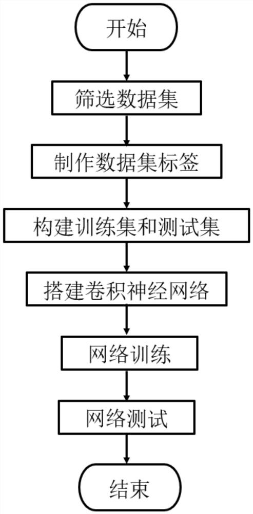
[0058] Embodiment 1: a kind of method of lesion segmentation, comprises steps:

[0059] S1, screen the data set and write the data interface required by the convolutional neural network according to the data characteristics of the data set;

[0060] S2, making data set labels;

[0061] S3, build training set and test set;

[0062] S4, the construction of convolutional neural network, the establishment of feature extraction model, input the image into the convolutional neural network, and obtain the segmentation results of multiple lesions respectively.

Embodiment 2

[0063] Embodiment 2: On the basis of Embodiment 1, in step S1, the data set includes the IDRiD data set and the DDR data set, and the data features include the lesion segmentation label and image resolution of the data set IDRiD and the data set DDR .

Embodiment 3

[0064] Embodiment 3: on the basis of embodiment 1, in step S2, comprise sub-step:

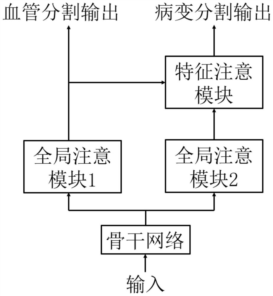
[0065] S21, Constructing a unified label for multi-lesion segmentation: Since different categories have different labeling labels, to achieve multi-category simultaneous segmentation, it is necessary to unify different labels on the same label, and use 0 to 4 to represent the background and four types of lesions respectively;

[0066] S22. Create pseudo-labels of blood vessels: Due to the high cost of medical image annotation, images with lesion annotations generally do not have pixel-level annotations of blood vessels. In the embodiment of the present invention, a segmentation model is pre-trained using the DRIVE and STARE data sets with blood vessel pixel level annotations, and the blood vessel pseudo-labels of the IDRiD and DDR data sets are obtained based on this model.


PUM


Abstract

The invention discloses a focus segmentation method and device and a medium, and belongs to the field of computer vision, and the method comprises a global attention module based on a transformer structure, a feature attention module based on the transformer structure, and the use of a cross entropy loss function with a weight based on the above modules. According to the method, detail information of the lesions and the blood vessels can be obtained, rich fundus prior information contained in blood vessel distribution can be fully utilized, the dependency relationship of different lesions in the image and the recessive pathological association between the lesions and the blood vessels can be obtained, and simultaneous segmentation of the DR multiple lesions can be more accurately achieved; the problem that foreground and background pixel points are unbalanced is solved, simultaneous accurate segmentation of multiple difficult and important lesions in actual medical diagnosis is achieved, and the technical problems that in the prior art, a segmentation model is difficult to learn standard features of the lesions, and wrong segmentation exists are solved.

Description

technical field [0001] The present invention relates to the field of computer vision, and more specifically, to a method, device and medium for lesion segmentation. Background technique [0002] The existing technical solutions for lesion segmentation have the following technical problems: 1. There are large differences between the same type of lesions in different disease development stages, making it difficult for the segmentation model to learn the standard features of lesions; 2. There are morphological differences between different lesions. A certain degree of similarity, such as hard exudate and soft exudate, makes the model appear mis-segmentation and mis-segmentation. Diabetic retinopathy (DR) will be described as an example below, but it is not limited to diabetes. [0003] Diabetic retinopathy (DR) is a complication caused by multiple long-duration diabetes. my country is the country with the largest number of diabetic patients in the world. The prevalence rate of...

Claims


Application Information

Patent Timeline
Patent Type & Authority Applications(China)
IPC IPC(8): A61B3/12
Inventor 许廷发黄诗淇李佳男肖予泽沈宁
Owner 北京理工大学重庆创新中心
Why Eureka
Try Eureka