
Method for enhancing echogenicity and decreasing attenuation of microencapsulated gases

A technology of gas and fluorine-containing gas, applied in echo/ultrasound imaging agents, preparations for in vivo tests, pharmaceutical formulations, etc., can solve problems such as non-standard harmonic imaging and no effort to correct the acoustic properties of ultrasound contrast agents

Inactive Publication Date: 2006-08-09
ACUSPHERE INC
View PDF10 Cites 0 Cited by
  • Summary
  • Abstract
  • Description
  • Claims
  • Application Information

AI Technical Summary

Problems solved by technology

However, harmonic imaging is currently not a standard
[0017] Importantly, these approaches do not address fundamental issues with the sonic properties of existing ultrasound contrast agents

Method used


Image

  • Method for enhancing echogenicity and decreasing attenuation of microencapsulated gases
  • Method for enhancing echogenicity and decreasing attenuation of microencapsulated gases
  • Method for enhancing echogenicity and decreasing attenuation of microencapsulated gases

Examples


Embodiment 1

[0094] Example 1: Preparation of polymer microparticles with enhanced echogenicity

[0095] 3.2g PEG-PLGA (75:25) (IV=0.75dL / g), 6.4g PLGA (50:50) (IV=0.4dL / g) and 384mg diarachidonic acid phosphatidylcholine were dissolved in 480ml di in methyl chloride. 20 ml of 0.18 g / ml ammonium bicarbonate solution was added to the polymer solution and the polymer / salt mixture was homogenized for 2 minutes using a Virtis homogenizer at 10,000 RPM. The solution was suctioned and spray dried with a BucchiLab spray dryer at a flow rate of 20ml / min, the inlet temperature was 40°C, and the outlet temperature was 20-22°C. Measured with a Coulter particle size analyzer, the particle diameter ranges from 1 to 10 microns, with an average of 2.0 microns. Scanning electron microscopy showed that the particles were generally spherical with a smooth surface and occasional surface shrinkage. Made into microspheres for transmission electron microscopy by embedding in LR white resin and then polymeriz...


PUM


Abstract

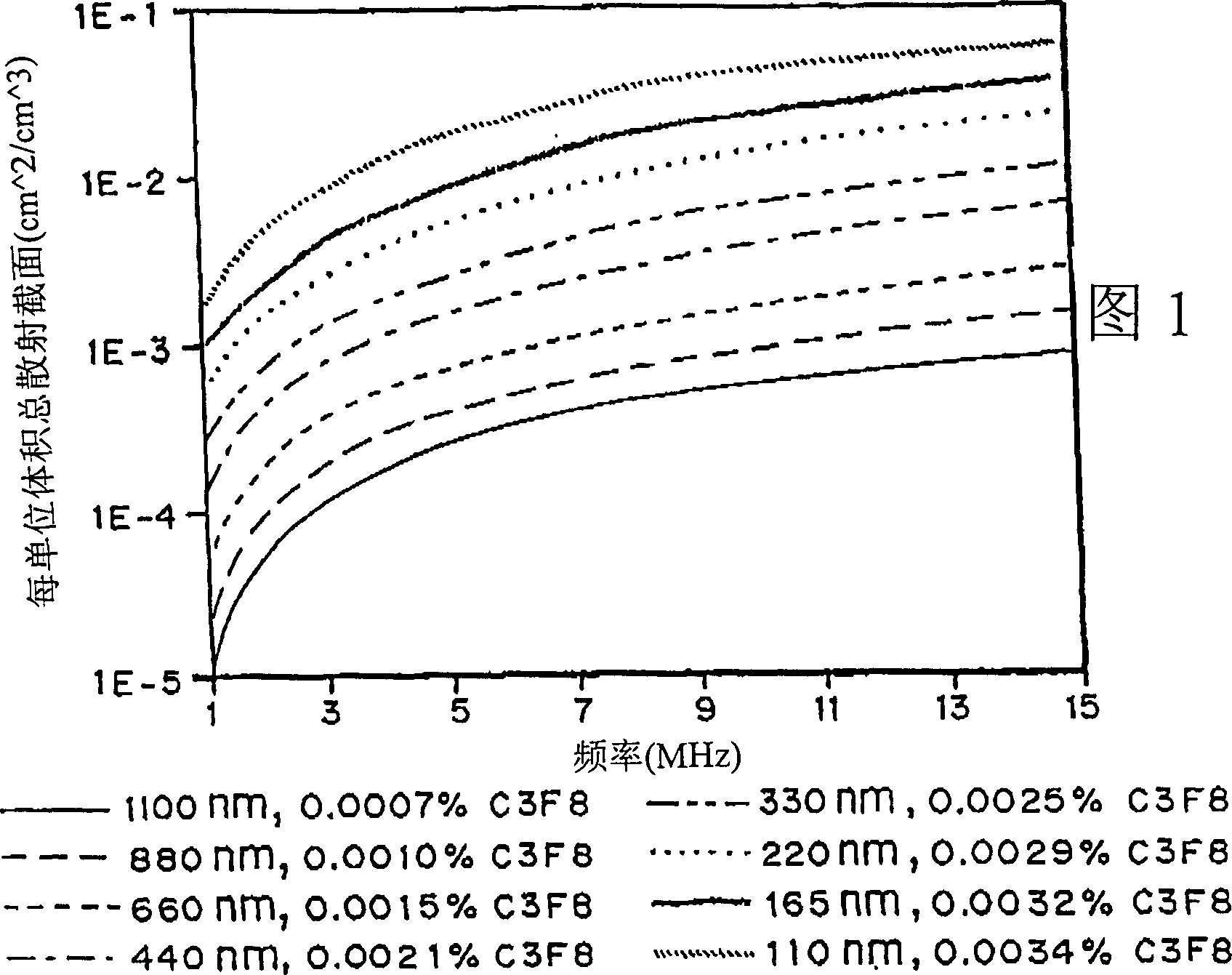
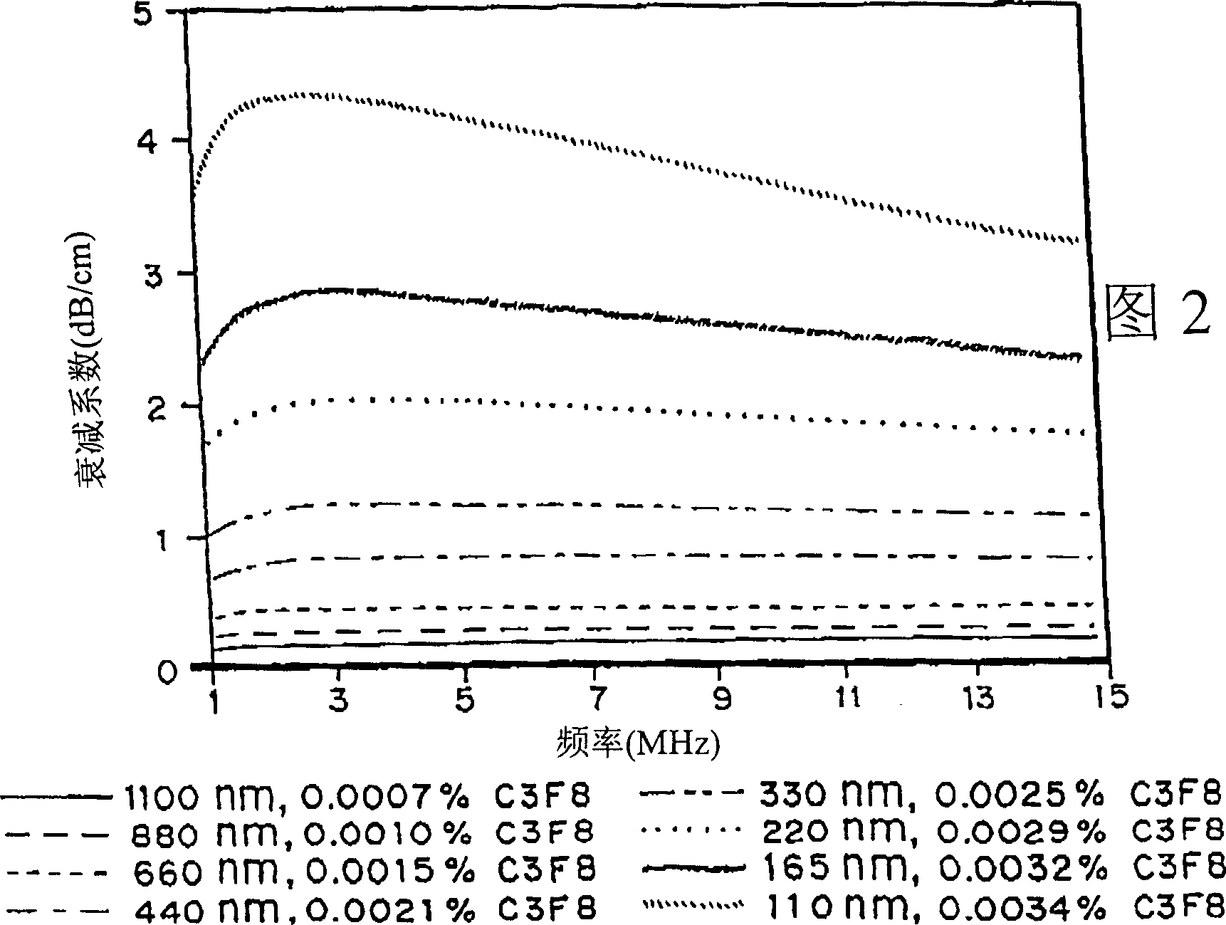
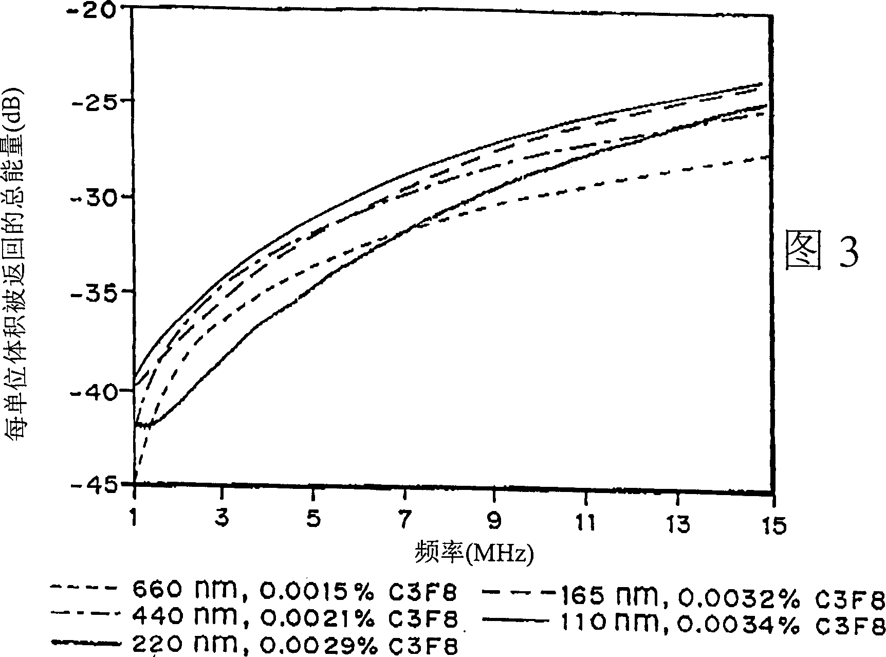
It has been discovered that microparticles formed from natural or synthetic polymer with thicker walls have significantly enhanced echogenicity as compared with microparticles having thinner walls. The effect of wall thickness has been determined experimentally as well as inserted into a formula for use in predicting the optimum conditions. In the preferred embodiment, the polymers are synthetic biodegradable polymers and the wall thickness is between about 100 and 660 nm, although wall thicknesses from about 20 nm to in excess to 500 nm can be used. The microparticles are manufactured with a diameter suitable for the targeted tissue to be imaged, for example, with a diameter for between 0.5 and 8 microns for intravascular administration, and a diameter of between 0.5 and 5 mm for oral administration for imaging of the gastrointestinal tract or other lumens. Preferred polymers are polyhydroxy acids such as polylactic acid-co-glycolic acid, polylactide or polyglycolide, most preferably conjugated to polyethylene glycol or other materials inhibiting uptake by the reticuloendothelial system (RES). The microspheres may be used in a variety of ultrasound imaging applications including cardiology applications, blood perfusion applications as well as for organ and peripheral vein imaging.

Description

Background of the invention [0001] The present invention is in the field of diagnostic imaging agents in general and, more particularly, to particulate ultrasound imaging contrast agents having greater echogenicity and less attenuation as a function of the thickness of the polymer film . [0002] When ultrasound is used to obtain images of internal organs or structures in a person or animal, ultrasound waves—those waves with frequencies higher than the human ear can detect—are reflected as they pass through the body. Different types of body tissues reflect ultrasonic waves differently, detect the reflections of ultrasonic waves produced by different internal structures, and convert them electrically into intuitive and visual images. [0003] For some medical diseases, it is very difficult to obtain useful images of target structures or organs, because in the absence of contrast-enhancing agents, ultrasound images produced by ultrasound reflections are not sufficient to distin...

Claims


Application Information

Patent Timeline
Patent Type & Authority Patents(China)
IPC IPC(8): A61K49/22A61B8/00A61K49/00
CPCA61B8/481A61K49/223A61K49/00
Inventor 查尔斯·C·丘奇霍华德·伯恩斯坦朱莉·安·斯特劳布亨利·T·布鲁什
Owner ACUSPHERE INC
Learn More