

Visible light induced delayed lighting no-damage tumor imaging method and equipment
A technology of delayed luminescence and tumor imaging, applied in the field of medical imaging diagnosis of tumors, can solve the problems of single optical characteristic difference of imaging contrast, difficult to accurately measure deep tissue, low imaging contrast, etc. high rate effect
- Summary
- Abstract
- Description
- Claims
- Application Information
AI Technical Summary
Problems solved by technology
Method used
Image

Examples
Embodiment 1
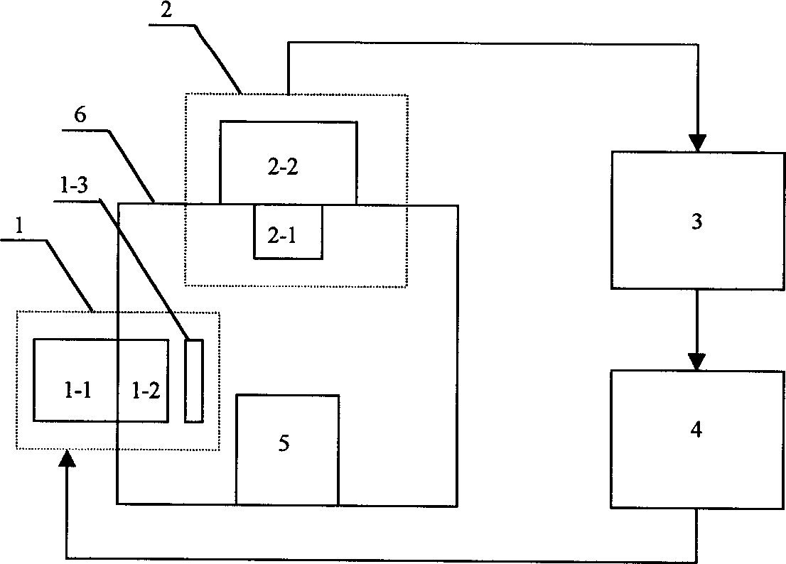
[0032] figure 1 shows the structure of the visible light-induced delayed luminescence non-damaging tumor imaging device of the present invention, by figure 1 It can be seen that the device includes a light excitation component 1, a light receiving component 2, an analog-to-digital converter 3, and a computer 4, wherein the light excitation component 1 is composed of an excitation light source 1-1, a focusing lens 1-2 and an optical filter group 1- 3; the light receiving component 2 is composed of a photo lens 2-1 that collects light and a detector 2-2; the light excitation component 1 is electrically connected to the computer 4, and the light receiving component 2, the analog-to-digital converter 3 and the computer 4 are in turn electrical connection; 5 is a three-dimensionally adjustable sample rack, on which the organism or tissue to be tested is placed, and 6 is a dark room. The device is composed of various components connected together, wherein the excitation light sourc...
Embodiment 2
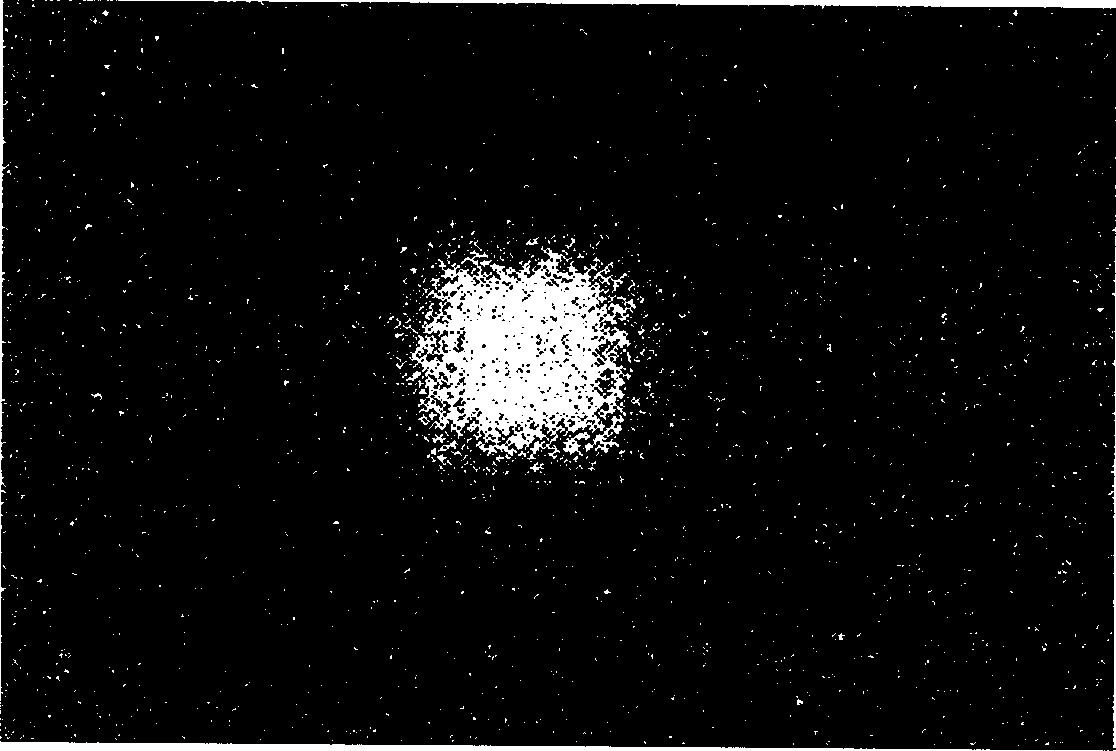
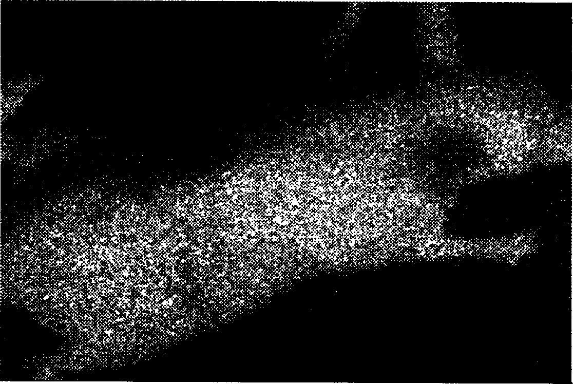
[0035] Apply the device described in Example 1 to the imaging of living organisms: take a 4-month-old experimental nude mouse, subcutaneously inoculate lung adenocarcinoma cells on the right shoulder of the nude mouse, two weeks later, the inoculation site grows about 1cm solid tumor. The day before the experiment, the nude mice were intravenously injected with the porphyrin photosensitizer chlorin e4 at a dose of 1-10 mg / Kg body weight, and 20-200 mg / Kg body weight of active serum albumin was subcutaneously injected 24 hours later. Nude mice were anesthetized, placed on the sample rack 5 in the darkroom 6, and first recorded under weak light as image 3 As shown in the outline drawing, locate the position of the mouse body and the tumor, then completely close the darkroom 6, and open the light-shielding shutter and power supply of the excitation light source 1-1 through the computer 4, so that the excitation light can irradiate the surface of the nude mouse vertically and uni...
PUM
 Abstract
Description
Claims
Application Information